


In this guide16 sections
- Key Points
- Hard Lump on Forehead That Feels Like Bone: Is It an Osteoma?
- What Causes an Osteoma on the Forehead?
- Forehead Osteoma Symptoms: Do They Cause Pain or Headaches?
- Is a Forehead Osteoma Dangerous? Can It Become Cancer?
- Forehead Lump: Osteoma vs Lipoma vs Cyst
- How Is a Forehead Osteoma Diagnosed? Do I Need a CT Scan?
- Do I Need to Do Anything About a Forehead Osteoma?
- Forehead Osteoma Removal: Endoscopic vs Open (Direct or Coronal) Surgery
- Am I Suitable for Endoscopic Forehead Osteoma Removal?
- Can a Forehead Osteoma Come Back After Removal?
- Forehead Osteoma Removal Before and After
- Forehead Osteoma Removal Recovery and Scarring
- Frequently Asked Questions About Forehead Osteoma
- Get a Forehead Lump Assessed in London
- References
Key Points
- A hard lump on the forehead that feels like bone and does not move is most often an osteoma, a slow-growing, benign bony growth on the frontal bone.
- A soft, squashy or mobile lump is usually something else, such as a lipoma or a cyst, and is treated differently.
- Most forehead osteomas need no treatment. Removal is a personal choice for appearance, or a medical one if the lump is growing, painful or involves the frontal sinus.
- A CT scan is the usual way to confirm the diagnosis and to plan surgery. Multiple osteomas, especially with a family history of bowel polyps, should prompt a conversation with your GP about Gardner’s syndrome.
- Endoscopic removal uses small cuts hidden behind the hairline and suits many smaller, well-defined lumps. Larger lumps, or those involving the frontal sinus, may need an open or combined approach.
- Recurrence is uncommon but has been reported, usually linked to bone left behind, so complete removal and histology matter.
Most people find it by accident. A hand across the forehead, a different angle in the mirror, and there it is: a hard bump that has probably been there for years. The reassuring answer first: in adults, a firm, fixed, painless lump on the forehead is very often an osteoma, a benign growth of bone. It does not spread and it is not a cancer. “Very often” is not “always”, though, and a few other lumps look similar, so it helps to know how to tell them apart. Our guides to the difference between a soft lump and a hard lump under the skin and lipoma vs cyst cover the wider picture.
This article explains how to recognise a forehead osteoma, what else it could be, how it is diagnosed, and how forehead osteoma removal works, including how to judge whether you are suitable for endoscopic (keyhole) removal rather than an open approach.
Hard Lump on Forehead That Feels Like Bone: Is It an Osteoma?
An osteoma is bone. That single fact produces most of what distinguishes it, and you can check it yourself in about thirty seconds.
- Press it. An osteoma is hard, not firm or rubbery. It feels like pressing your own skull, because you largely are.
- Try to move it. Push it from side to side. An osteoma does not move, because it is attached to the bone beneath. The skin slides over it; the lump stays put.
- Check whether it hurts. Most are painless. Tenderness is possible, but pain is not the typical picture.
- Think about how fast it appeared. Osteomas grow slowly, over years. A hard lump that appeared over weeks deserves a closer look.
- Feel the shape. Usually round or oval, smooth or slightly bumpy, with a defined edge.
Hard, fixed, painless and slow-growing on the forehead is a characteristic combination. In a 2026 Korean review of 148 craniofacial osteomas, the frontal bone was the most common site, accounting for about 60% of cases[1]. If you searched for a hard lump on the forehead under the skin, a hard bony lump on the forehead or a hard lump on the forehead above the eyebrow or near the hairline, the same test applies wherever the lump sits.
What Causes an Osteoma on the Forehead?
In most people, nobody knows. For a single osteoma with no family history, no specific cause is usually identified. Several possibilities have been proposed, none of them proven for every case:
- Previous injury. Some people recall a knock to the same spot years earlier. Whether the injury triggered the growth or simply drew attention to one already forming is unclear.
- Inflammation of the covering of the bone.
- Developmental variation. Some osteomas seem to be present from early life.
- Genetics. Rarely, multiple osteomas are part of an inherited condition (see Gardner’s syndrome below).
Reports differ on who is affected. A large Korean series found most patients with craniofacial osteomas were women[1], whereas studies of osteomas inside the frontal sinus describe a male predominance[7]. Osteomas are often noticed in adulthood simply because they are slow to grow and easy to overlook.
Forehead Osteoma Symptoms: Do They Cause Pain or Headaches?
Most forehead osteomas cause no symptoms at all beyond a visible or palpable bump. Case series note that they are usually silent until they become large[2]. Symptoms are more likely when the bone growth sits over a nerve, presses on nearby structures, or grows from the frontal bone into the frontal sinus or towards the eye socket. Persistent localised headache, tenderness, a blocked or infected sinus, or changes in vision or eye position are not typical of a simple surface osteoma and are reasons to be assessed.
Is a Forehead Osteoma Dangerous? Can It Become Cancer?
A typical forehead osteoma is not dangerous. Osteomas are not known for malignant transformation, and recurrence after removal is described as rare[3]. The caution is that a lump that grows quickly, hurts, or changes the overlying skin is behaving differently from a classic osteoma, and a bone tumour that is not an osteoma needs to be excluded rather than assumed away.
Multiple osteomas and Gardner’s syndrome
A single osteoma is almost always an isolated finding. Multiple osteomas are a different matter, particularly alongside a family history of bowel polyps or colon cancer. Gardner’s syndrome is a form of familial adenomatous polyposis in which skull osteomas appear together with colonic polyps and other features, and it carries a high risk of bowel cancer[4]. The osteomas themselves are harmless; the bowel findings are not. Reports describe the bone and dental changes appearing before any bowel symptoms[5], which is why more than one bony lump on the head, or a family history of polyps, is worth mentioning to your GP whether or not you want anything done cosmetically.
Forehead Lump: Osteoma vs Lipoma vs Cyst
Not every forehead lump is an osteoma. The main alternatives are below, and the diagram sets them side by side.
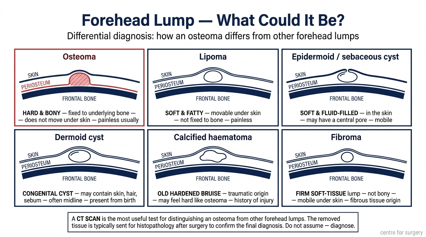
Lipoma (soft, movable). A benign fatty lump that sits in the tissue above the bone, so it is soft and moves when you push it. Soft and mobile points to a lipoma; hard and fixed points to an osteoma. Our guide to what a lipoma feels like describes the difference in more detail, and lipoma removal is a different and simpler procedure.
Epidermoid or sebaceous cyst. A lump within the skin itself. It feels softer, sometimes slightly springy, may have a tiny central pore, and moves with the skin. See sebaceous cyst vs epidermoid cyst. Higher up, in the hair-bearing scalp, a firm cyst is more often a pilar cyst.
Dermoid cyst. Usually present from early life, often near the midline or the outer end of the eyebrow.
Calcified haematoma. An old bruise that has hardened. It can feel very like an osteoma, and the clue is a definite injury to that exact spot.
Fibroma. Firm but made of soft tissue rather than bone, and usually has some mobility.
Other bone lesions. Uncommon, but the reason a bony lump should not be removed without knowing what it is. In one endoscopic series of 36 forehead tumours, the removed tissue turned out to be 18 lipomas, 13 osteomas, two dermoid cysts and one bone fragment, which shows how often lumps are not what they were assumed to be[6].
A lump can also be bone-hard because of the shape of your skull rather than any growth, which is why a prominent brow is sometimes mistaken for a lump. If the whole brow ridge is prominent rather than one spot, see how to reduce a brow bone and our guide to a big forehead.
How Is a Forehead Osteoma Diagnosed? Do I Need a CT Scan?
Examination alone is often enough for an experienced clinician to be fairly confident: hard, fixed, painless and slow-growing on the frontal bone is a recognisable picture. Imaging is what settles the question and shapes any operation. In the Korean series, every patient had a 3D CT scan for diagnostic confirmation[1].
A CT scan confirms the lump is bone rather than soft tissue, shows its exact size and depth, and shows whether it is confined to the outer surface of the frontal bone or extends towards the frontal sinus. That distinction changes the surgical approach. Plain skull X-rays show less detail and have largely been replaced by CT for this purpose. A biopsy is rarely needed beforehand where the CT appearance is characteristic, and removed tissue is normally sent for histopathology afterwards to confirm the diagnosis.
Do I Need to Do Anything About a Forehead Osteoma?
Usually not. A small, stable, painless osteoma that does not bother you can be left alone. Case series describe surgery as appropriate when the lesion is symptomatic or actively growing[2], and for appearance when it matters to the patient. Osteomas are bone, so they do not shrink or disappear on their own, and there is no evidence that creams, supplements or home remedies dissolve them. If you searched for a natural treatment or forehead osteoma removal without surgery, the honest position is that removal is a surgical procedure.
That is a personal decision, and nobody should feel pressured into removing a harmless bump. If you do want it dealt with, forehead contouring surgery may be relevant where the forehead shape more broadly is the concern, and a consultation will establish which option applies.
Forehead Osteoma Removal: Endoscopic vs Open (Direct or Coronal) Surgery
There are three broad ways to remove a forehead osteoma:
- Direct (open) excision: a cut directly over the lump, or in a natural forehead crease, to expose and remove it.
- Endoscopic removal: one or more small cuts hidden behind the hairline. A thin camera (endoscope) is passed beneath the scalp so the surgeon can see the bone on a screen and remove the lump with fine instruments.
- Coronal (open) approach: a longer cut across the scalp, from ear to ear, behind the hairline. It gives wide access and is reserved for larger or more complex lumps.
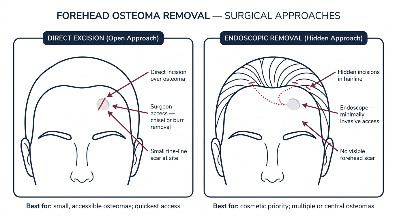
The endoscopic approach has been described in forehead surgery for over a decade. A 2014 multicentre retrospective study in JAMA Facial Plastic Surgery reported 36 patients who had benign forehead tumours removed endoscopically ↗. Complete excision was achieved in all, the mean operating time was 36 minutes, and there were no haematomas, infections, scalp numbness or recurrences; one patient had a patch of hair loss that resolved on its own[6]. That is encouraging, but it is a modest case series, and not every lump in it was an osteoma.
The wider evidence on osteomas in the frontal sinus points the same way. A 2019 systematic review of 1,399 patients ↗ found postoperative complications in 7.5% of endoscopic cases compared with 22.3% of open or combined cases, with a shorter hospital stay after endoscopic surgery[7]. Those are sinus osteomas, which differ from surface osteomas of the forehead, and the groups were not matched, so the figures show a trend rather than a promise for any individual.
Surgeons continue to refine the technique with case reports describing removal through two small hairline cuts[8], a mini-trocar to protect soft tissue from the burr[9], and a single incision about 2 cm behind the hairline[10]. In practice, the choice comes down to the lump and the surgeon rather than a rule that one method is always better.
Am I Suitable for Endoscopic Forehead Osteoma Removal?
Suitability is decided at a face-to-face assessment with imaging. These are the factors that matter most.
Size and position of the osteoma
Endoscopic removal is generally considered for small to medium, well-defined osteomas on the outer surface of the forehead that can be reached from behind the hairline. Lumps close to the midline or in the central forehead are straightforward to reach; those very near the eyebrow, temple or orbit rim need more careful planning. For large osteomas, a review of giant fronto-ethmoidal lesions found that size alone did not decide the approach, and that the choice rested on surgeon experience, available equipment and the anatomy involved[11].
When endoscopic removal is not suitable
An open or combined approach is more likely to be advised when:
- the CT shows the osteoma extends into the frontal sinus, towards the eye socket or through the inner bone. In the 2019 sinus review, 18.7% had extended into the orbit and 9.5% inside the skull[7], so these cases belong with a specialist sinus or craniofacial team;
- the lump is very large, or very broad-based, so that it cannot be shaved down safely through small ports;
- there are several osteomas across a wide area;
- the diagnosis is uncertain, and a wide view or tissue sample is needed first.
Scar and hairline position
Endoscopic cuts sit behind the hairline, so they are hidden by hair, whereas a direct cut sits on the forehead and leaves a visible line, though a scar placed in a forehead crease may blend in better. A coronal cut is long but also hidden in the hair. If your hair is very short or thinning, or you wear your hair back, discuss exactly where each scar would fall. Our guide to endoscopic vs traditional brow lift explains the same hidden-incision principle used in forehead surgery generally.
Recovery and general health
The approach affects swelling, how many dressings and how soon you feel presentable, but healing is individual. Smoking, conditions that slow wound healing and blood-thinning medication all need to be discussed beforehand, and your surgeon will tell you what to stop and when. Step-by-step recovery guidance is set out on the forehead osteoma removal page.
What happens at the consultation
The surgeon examines the lump, reviews your history including any family history of bowel polyps, and checks the CT scan. You will discuss what you want to change, which approach suits the anatomy, where the scars would sit, the anaesthetic (local anaesthetic with sedation, or TIVA, depending on the case) and the risks. If a scan has not been done, one is usually requested before a decision is made. If the diagnosis turns out to be a lipoma or cyst, you will be directed to the right procedure instead.
Can a Forehead Osteoma Come Back After Removal?
Recurrence is uncommon, but it is not impossible, and it is not honest to say there is no risk. Where recurrence has been reported, it has been linked to bone left behind. A 2023 case report in the World Journal of Clinical Cases ↗ described a woman whose forehead osteoma returned after two earlier endoscopic removals; the authors suggested that residual fragments of osteoma may have seeded the surface, and advised meticulous burring and a thorough inspection of the bone surface after irrigation[12]. In the sinus systematic review, complete resection was a significant predictor of whether disease recurred or progressed[7], and other series describe recurrence as rare[3].
The practical message is that a careful, complete removal with the tissue checked by a pathologist gives a good chance of a lasting result, and that you should return for review if a lump reappears.
Forehead Osteoma Removal Before and After
These photographs show forehead osteoma removal at Centre for Surgery. Results vary between patients and depend on the size and position of the lump.


Forehead Osteoma Removal Recovery and Scarring
After removal you can expect a dressing, some swelling and bruising, and tenderness around the area; the extent varies with the approach and the size of the lump. Temporary numbness of the skin near the scar is possible whichever approach is used. Your surgeon will give written aftercare advice, including how long to avoid strenuous activity and how to protect the area from knocks and sun while it heals. In the endoscopic series above, no patient had scalp numbness or contour irregularity[6], but that is one group of patients and should not be treated as a guarantee. Scars mature over months, and your surgeon can advise on scar care. If what you really want is a smoother forehead overall rather than removal of one lump, a brow lift or endoscopic brow lift is a different operation for a different problem, and a consultation will help sort out which you need.
Frequently Asked Questions About Forehead Osteoma
Why do I have a hard bump on my forehead?
The most likely explanation is an osteoma, a benign, slow-growing bone growth on the frontal bone. These are hard, fixed in place and usually painless. Other possibilities include a lipoma, a cyst or a calcified old bruise. A lump that is genuinely hard and does not move is most often an osteoma, but only examination, and sometimes a scan, can confirm it.
What is a calvarial osteoma?
The calvaria is the domed top of the skull, which includes the forehead. A calvarial osteoma is a benign bony growth that arises from the outer or, less often, the inner surface of those skull bones. A forehead osteoma is the most familiar example.
Is an osteoma of the forehead a serious problem?
Usually not. A typical forehead osteoma is benign and is not known to turn cancerous. It becomes a concern if it grows quickly, causes pain, involves the frontal sinus or eye socket, or if there are several of them, because multiple osteomas can be linked with Gardner’s syndrome. Those situations warrant assessment.
Does an osteoma ever stop growing?
Osteomas grow slowly, often so slowly that the change is hard to notice, and some stay the same size for years. There is no reliable way to predict which will keep growing, so a lump that is measurably getting bigger, or is changing quickly, should be examined.
Can a forehead osteoma affect the brain?
A typical osteoma on the outer surface of the forehead sits outside the skull and does not press on the brain. Osteomas of the frontal sinus are different: in one systematic review, about one in ten had extended inside the skull[7]. A CT scan shows which type you have.
What is the difference between an osteoma and a lipoma?
Consistency and movement. An osteoma is bone: hard and fixed to the skull. A lipoma is fat: soft, squashy and movable under the skin. If you can push it around, it is unlikely to be an osteoma. They also need different procedures, because a lipoma is removed through the soft tissue while an osteoma needs the bone surface reshaped. See our guide to whether lipomas come back for more on that side.
Will a forehead osteoma go away on its own?
No. An osteoma is mature bone, so it does not shrink or dissolve, and no cream, supplement or home remedy has been shown to remove one. Left alone, it will usually stay as it is or grow very slowly.
Can a forehead osteoma be removed without surgery?
No non-surgical treatment has been shown to remove bone. Removal means surgery, either endoscopic through small hairline cuts or by an open approach. If nothing is bothering you, monitoring is a reasonable option.
Does the NHS remove forehead osteomas?
Usually only when there is a clear medical reason, such as symptoms, growth or sinus involvement. Removal for appearance alone is generally treated as cosmetic and is not routinely funded, though policies vary by area. Ask your GP what applies where you live.
Can a forehead osteoma come back after removal?
Recurrence is uncommon but has been reported, mostly where bone was left behind. Complete removal, careful finishing of the bone surface and histology reduce the chance, and you should return for review if a lump reappears.
Do I need a scan for a forehead osteoma?
Not always to suspect one, because examination is often suggestive. A CT scan is the usual next step if removal is being considered, if the diagnosis is unclear, or if there are symptoms, because it shows the size, depth and sinus involvement that determine how surgery is done.
Get a Forehead Lump Assessed in London
If you are unsure what your forehead lump is, the first step is examination by someone who assesses them regularly. That will usually settle the question, and the honest answer is often that nothing needs doing. Centre for Surgery is regulated by the Care Quality Commission, and consultations take place at 95–97 Baker Street, Marylebone.
Phone: 020 7993 4849
Email: contact@centreforsurgery.com
Contact us to arrange an assessment
References
- Moon JE, Kang HS, Chang YJ, et al. Diagnostic and treatment protocols for peripheral craniofacial osteomas. Journal of Craniofacial Surgery 2026. doi:10.1097/SCS.0000000000012685 ↗
- Gundewar S, Kothari DS, Mokal NJ, Ghalme A. Osteomas of the craniofacial region: a case series and review of literature. Indian Journal of Plastic Surgery 2013;46(3):479–485. doi:10.4103/0970-0358.121982 ↗
- Nguyen S, Nadeau S. Giant frontal sinus osteomas: demographic, clinical presentation, and management of 10 cases. American Journal of Rhinology & Allergy 2019;33(1):36–43. doi:10.1177/1945892418804911 ↗
- Andour H, Mamouch A, Hassar S, et al. Gardner syndrome: when cervical-facial osteomas reveal the tip of the iceberg. Radiology Case Reports 2025;20(4):1967–1971. doi:10.1016/j.radcr.2024.12.043 ↗
- Yu D, Ng BCW, Zhu H, Liu J, Lin Y. Bone and dental abnormalities as first signs of familial Gardner’s syndrome in a Chinese family. Médecine/Sciences 2018;34(F1):20–25. doi:10.1051/medsci/201834f104 ↗
- Sadick H, Huber M, Perkins SW, et al. Endoscopic forehead approach for minimally invasive benign tumour excisions. JAMA Facial Plastic Surgery 2014;16(5):352–358. doi:10.1001/jamafacial.2014.269 ↗
- Karunaratne YG, Gunaratne DA, Floros P, Wong EH, Singh NP. Frontal sinus osteoma: from direct excision to endoscopic removal. Journal of Craniofacial Surgery 2019;30(6):e494. doi:10.1097/SCS.0000000000005371 ↗
- Raponi I, Giovannetti F, Cacciatore F, Di Carlo D, Valentini V. Endoscopic forehead approach for minimally invasive frontal osteoma excision. Journal of Craniofacial Surgery 2020;31(8):e767–e769. doi:10.1097/SCS.0000000000006715 ↗
- Chen S, Deng Y, Xu Y, et al. Endoscopic approach with an innovative mini-trocar for forehead osteoma excision. Journal of Craniofacial Surgery 2024;35(2):e154–e156. doi:10.1097/SCS.0000000000009921 ↗
- da Costa MDS, Suzuki FS, Biló JP, Cavalheiro S. Endoscopic approach for resection of frontal forehead osteoma: technical case report instruction. World Neurosurgery 2023;175:11. doi:10.1016/j.wneu.2023.03.135 ↗
- Humeniuk-Arasiewicz M, Stryjewska-Makuch G, Janik MA, Kolebacz B. Giant fronto-ethmoidal osteoma: selection of an optimal surgical procedure. Brazilian Journal of Otorhinolaryngology 2018;84(2):232–239. doi:10.1016/j.bjorl.2017.06.010 ↗
- Lee DY, Lim S, Yoon JS, Eo S. Recurred forehead osteoma disseminated after previous osteoma excision: a case report. World Journal of Clinical Cases 2023;11(31):7684–7689. doi:10.12998/wjcc.v11.i31.7684 ↗