




Pyogenic granuloma removal in London: excision and laser
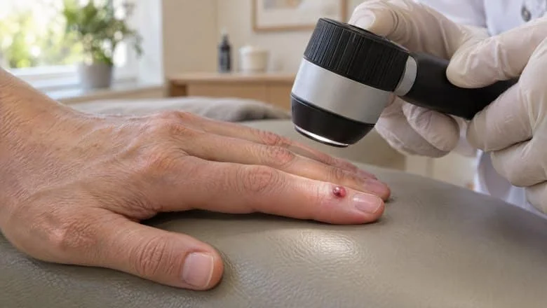
Pyogenic granuloma removal treats a small, bright red lump that appears over a few days or weeks and bleeds at the slightest knock. Its medical name is lobular capillary haemangioma: a harmless overgrowth of tiny blood vessels, most often on a finger, around a nail, on the lip or on the face. It cannot turn into cancer, but it bleeds again and again, catches on clothing and, outside pregnancy, rarely goes away on its own.
At Centre for Surgery, our usual pyogenic granuloma treatment is surgical excision: the lump is cut out under local anaesthetic, the wound is closed with fine stitches and the tissue is sent for histology. Excision is carried out by a GMC-registered consultant plastic surgeon at our Baker Street hospital in Marylebone. For a small, superficial lesion with a clear diagnosis, laser on the Fotona SP Dynamis Pro is an alternative that avoids stitches but leaves no tissue to examine. One of our plastic surgeons examines the lump first to confirm it is suitable for laser, and Ida Bafende, NMC-registered aesthetic practitioner, then carries out the laser treatment.
That trade-off matters because a few rarer growths can look like a pyogenic granuloma, most importantly a melanoma that makes no dark pigment. Below, we explain how they are told apart, which treatment suits which lump and the healing time by body site. A separate page describes how skin lesions are assessed and removed in general.
A pyogenic granuloma is one of many lumps people ask us about. Our plastic surgeon’s guide to common skin lumps and bumps explains the others, and you can see everything we treat under lumps and bumps removal.
Pyogenic granuloma removal at a Glance
Laser is only offered for a small, superficial lump that one of our plastic surgeons has assessed as clearly benign; anything uncertain is cut out and sent for analysis, and treatment waits until after pregnancy and breastfeeding.
Considering pyogenic granuloma removal?Book a consultation to have your lump assessed and to find out whether excision or laser suits it. No obligation, and no pressure to book.
Pyogenic granuloma removal before and after photos
The photo below shows a pyogenic granuloma before and after laser treatment, which is only used once the lump has been assessed as suitable.
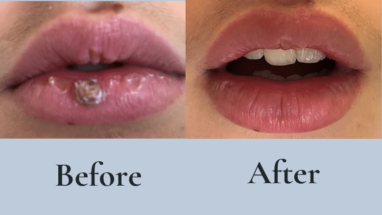
Results depend on the size, depth and site of the lump. Laser can need more than one session, a lump that is cut out heals with a fine line scar, and any pyogenic granuloma can grow back, so your clinician talks you through what to expect for your own lesion.
What is a pyogenic granuloma (lobular capillary haemangioma)?
A pyogenic granuloma is a benign growth of small blood vessels (capillaries) in the skin, or sometimes in the lining of the mouth or nose. Doctors also call it a lobular capillary haemangioma (hemangioma in American spelling), which describes it better: under the microscope it is made of lobules, or clusters, of capillaries. The older name is misleading: “pyogenic” means pus-forming, but it is not caused by infection, and it is not a true granuloma.
What does a pyogenic granuloma look like?
- Colour: bright red to purple-red, often compared to a raspberry.
- Size: usually 5–10 mm across, occasionally larger.
- Surface and shape: a soft, glistening dome, sometimes on a stalk, often crusted and with a collar of thickened skin at its base.
- Growth: it appears suddenly and grows quickly over days to weeks.
- Bleeding: a light knock or scratch can start bleeding that is hard to stop.
The thin surface stretched over so many small vessels is why it bleeds so readily, and why the feeding vessel at its base is dealt with when it is removed.

It is not the same as the strawberry-like haemangioma some babies develop, which grows for months and then slowly shrinks by itself.
Where do pyogenic granulomas occur?
They can appear almost anywhere, but in a UK audit of 408 cases the head and neck was the commonest area[1]. Typical sites are the fingers and hands (especially the skin fold beside a nail), the lips, the face, nose, eyelids and scalp, the gums and inside of the mouth in pregnancy, the upper body and the feet. They are commonest in children and young adults and in pregnancy, and some medicines make them more likely. The British Association of Dermatologists’ patient guide to pyogenic granulomas is a clear independent summary.
What causes a pyogenic granuloma?
The exact cause is not fully understood. A pyogenic granuloma seems to form when the normal repair of the skin overshoots, so that new blood vessels laid down during healing keep growing.
Common triggers
- Minor skin injury: a scratch, splinter, insect bite, burn or an ingrown nail, often a few weeks before the lump appears.
- Piercings: a red lump that bleeds beside a nose or ear piercing is often a small pyogenic granuloma; our guide to piercing bumps covers these separately.
- Pregnancy and hormonal change: especially on the gums and lips. These often settle after the baby is born.
- Medicines: retinoid tablets used for severe acne, some chemotherapy drugs, some antiretroviral medicines and oral contraceptives have all been linked to pyogenic granulomas.
- Healing wounds and burns: overgrown healing tissue in a wound can turn into a pyogenic granuloma.
- No trigger at all: a large share appear spontaneously.
A pyogenic granuloma is not caused by poor hygiene, is not contagious and is not an infection, although a raw, bleeding surface can become infected.
Pyogenic granuloma vs amelanotic melanoma: why histology matters
Most pyogenic granulomas are easy to recognise, but a few growths can look almost identical, and one is a skin cancer. Amelanotic melanoma makes little or no dark pigment, so it can look like a pink or red lump that bleeds rather than a dark mole; read more about how melanoma differs from a benign lesion. A dermatoscope (a magnifying light held against the skin) helps, but a 2010 multicentre study of 122 pyogenic granulomas and 140 other growths concluded that dermoscopy is not a substitute for histology, because melanoma cannot be ruled out when vessels are visible[2].
Why the tissue goes to the laboratory
Tissue that is cut out (an excision, shave, punch or biopsy) is sent to an accredited laboratory for histology, where a pathologist examines it under the microscope. A 2007 UK audit of 408 pyogenic granulomas in the Journal of Plastic, Reconstructive & Aesthetic Surgery (Giblin et al.) put it plainly: whatever technique is used, it must yield material for analysis so that other diagnoses can be excluded[1]. That is why laser, which destroys the tissue, is kept for small lesions with a clear diagnosis. Results usually take 3–4 weeks, and we contact you with the result; our guide to what your histology report means explains what it says.
The same applies to children. In a 2025 single-centre series of 84 children whose lump was thought to be a pyogenic granuloma, 6 turned out to be a Spitz naevus once the tissue was examined[3]. A Spitz naevus is a type of mole, and our mole removal page covers how moles are treated.
Other lumps that can look like a pyogenic granuloma
- Cherry angioma: a smaller, smooth, evenly red spot that develops slowly and rarely bleeds; see cherry angioma removal and our article on red moles and other red spots.
- Glomus tumour and eccrine poroma: rare benign growths; a glomus tumour is usually under a nail and very painful.
- Kaposi sarcoma and bacillary angiomatosis: purple-red lumps, usually several, seen mainly when the immune system is weakened.
- Other skin cancers: occasionally a squamous cell carcinoma, or very rarely an angiosarcoma, mostly in older people.
When we take extra care
Age over 40, sun-damaged skin, slower growth over months, pigment around the edge or unusual dermoscopy features mean the lump is cut out whole and sent for analysis rather than lasered. If your immune system is weakened by medicines, a transplant or HIV, a sample is examined before any treatment that destroys tissue, and we liaise with the team that looks after you where appropriate. We do not treat skin cancer at Centre for Surgery. If a lump looks as though it could be a skin cancer, or a biopsy shows one, we refer you through your GP to an NHS skin cancer team, on the urgent suspected cancer pathway where that applies.
Do I need to have a pyogenic granuloma removed?
You do not have to, but most people choose to, because outside pregnancy a pyogenic granuloma rarely goes away by itself. The usual reasons are:
- Repeated bleeding: the commonest reason; a minor knock can stain clothing or bedding.
- It gets in the way: a finger lump catches on things, and a lip lump interferes with eating and speaking.
- Appearance: a red lump on the face, lip or hand is hard to hide.
- Peace of mind: confirming the diagnosis rather than living with a lump nobody has examined.
- A smaller scar: a small lump removed early generally leaves a smaller mark.
When to get it checked sooner
Be seen promptly if the bleeding is heavy or hard to control, an eyelid lump affects your vision, you have a bleeding disorder or take a blood thinner, you are over 40, the lump is on sun-damaged skin or looks unusual, or it grows fast while your immune system is weakened. Our article on when a skin lump needs removing explains why fast growth and bleeding deserve a prompt look; the same goes for a bleeding mole.
If you take a blood thinner, tell us at your consultation and do not stop it unless the doctor who prescribed it advises you to. If you take aspirin that was not prescribed for you, stop it 2 weeks before your procedure; if your doctor prescribed it, ask them first.
If it is bleeding now, or you would rather wait
Press firmly on the lump with a clean cloth for 10 minutes without lifting it; if it is still bleeding heavily after 20 minutes, go to an urgent treatment centre or A&E. Leaving a lump is reasonable if it is very small and not bleeding, or if it appeared in pregnancy and may settle after the birth; have it assessed if it changes, grows or starts to bleed.
Pyogenic granuloma treatment: surgery, laser and other options
Pyogenic granuloma treatment aims to remove the whole lump, including the feeding vessel at its base, so that it stops bleeding and is less likely to grow back. Everything is done under local anaesthetic in our day-case procedure rooms; for a finger or toe, a ring block (anaesthetic injected at the base of the digit) numbs the whole digit, and you stay awake throughout.
Your consultation and assessment
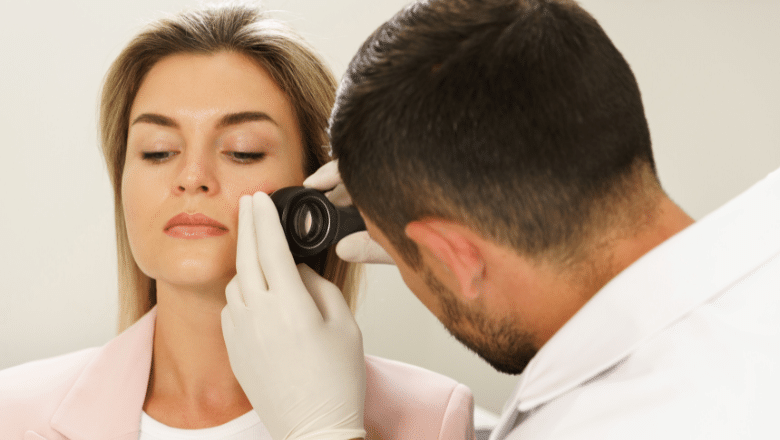
Your consultation takes about 30 minutes. One of our plastic surgeons asks about the lump and your health, examines it, with a dermatoscope where needed, and explains the likely diagnosis and the recommended technique. Before any laser treatment, the surgeon confirms the lump is benign and suitable for laser. If the lump does not look typical, a small sample (biopsy) may be taken first, or the whole lump removed for analysis.
We recommend a two-week cooling-off period between your consultation and treatment. If you have had a virtual consultation first and sent us photos of the lump, treatment can take place at the same visit as your in-clinic assessment, as long as the surgeon (or Ida, for laser) is happy it is suitable to treat on the day. Our guide to same-day removal explains what to expect.
Pyogenic granuloma surgery (excision)
Excision is our usual approach, and it is carried out by a consultant plastic surgeon. The lump is removed in a small ellipse of skin down to its base, including the feeding vessels, which are tied or sealed with heat to control bleeding. The wound is closed with fine stitches, planned along the natural creases of the skin where possible. It takes 20–30 minutes, or 30–45 minutes for a larger or recurrent lump.
Excision removes the whole lesion in one session for most people, gives the laboratory the complete lump to examine, and in the published evidence has the lowest regrowth rate of the surgical methods[4]. The trade-off is a fine line scar, a little longer than the lump because the skin is closed in a straight line. Our article on why every specimen goes to the lab explains the thinking behind sending tissue for analysis.
Pyogenic granuloma laser treatment
For a small, superficial pyogenic granuloma on the face or another visible area, where the diagnosis is clear, laser is an alternative. Once one of our plastic surgeons has confirmed the lump is suitable, Ida Bafende carries out the treatment with two wavelengths of the Fotona SP Dynamis Pro: the long-pulse Nd:YAG (1064 nm) seals the blood vessels feeding the lump, and the Er:YAG (2940 nm) can then remove the surface in thin layers with little heat. There are no stitches; a small scab forms and comes away as the skin heals.
The evidence is encouraging but limited. A 2012 prospective series of 20 patients in Dermatologic Surgery (Hammes et al.) used the long-pulse Nd:YAG laser: 19 of the 20 healed without regrowth over at least 6 months, with slight or no change in skin texture, but some needed up to four sessions[5]. It was a small study without a comparison group, so expect that more than one session may be needed.
Laser destroys the tissue, leaving nothing for the laboratory to examine, so it is not used when the diagnosis is unclear, you are over 40 and amelanotic melanoma is a possibility, the lump has unusual features, is large or deep, or has come back after previous laser. A lump whose nature is uncertain at your assessment is not lasered: it is removed surgically by one of our plastic surgeons and sent for analysis.
Curettage and cautery, freezing and other treatments
The British Association of Dermatologists describes curettage and cautery, where the lump is scraped off and its base sealed with heat, as the usual treatment, and the scrapings can be sent for analysis. Freezing (cryotherapy) is also used: in a 2006 randomised trial in which 76 patients completed treatment, every lesion cleared with either method, but freezing needed more sessions on average[6]. Silver nitrate cautery has also been reported[4], and DermNet lists further options, including prescribed beta-blocker gels such as timolol.
At Centre for Surgery, the treatments offered for pyogenic granuloma are excision and, for selected lesions, laser.
Pyogenic granuloma on the finger, nail, lip and face
Pyogenic granuloma on the finger or around the nail
Fingers and toes are common sites, often beside the nail after an ingrown nail or nail injury. A lump in the nail fold can regrow if the nail problem continues, so nail care is part of your recovery advice. One under the nail plate needs part of the nail removing first, and a fingernail then takes 3–6 months to grow back. Hand stitches stay in for 10–14 days. Two lookalikes matter here: warts around the nail, which are rough and skin-coloured, and glomus tumour, which is usually very painful.
Pyogenic granuloma on the lip
The lower lip is a common site, after a bite or in pregnancy. Lip tissue heals quickly and stitches usually come out at 5–7 days, but the edge of the red part of the lip needs careful alignment so that the lip line heals evenly. A soft, dark blue or purple bump that does not bleed easily is more likely a venous lake; other lip lumps, such as mucoceles, are covered under lip lesion removal.
Pyogenic granuloma on the face, nose and eyelid
On the face, the incision follows the natural lines of the skin, and laser is most often considered here for a small, superficial lesion. A lump on the outer skin of the nose is treated like any facial lesion, but one inside the nostril needs an ear, nose and throat (ENT) assessment. Lesions on the eyelid margin itself are removed by one of our facial plastic surgeons, usually by excision under local anaesthetic; a lump on the inner lining of the eyelid or on the eye itself needs an eye doctor (ophthalmologist).
In the mouth, on the scalp and on the body
Gum lumps, sometimes called a pregnancy tumour (epulis gravidarum), are common in pregnancy and often settle after the birth. Lumps on the gums or inside the mouth are usually managed by a dentist or oral surgeon, so we advise you on the right route and liaise with your dentist where needed. Scalp stitches usually come out at 5–7 days; on the body, arms and legs, at 10–14 days.
Pyogenic granuloma in pregnancy and in children
Pyogenic granuloma in pregnancy
Pregnancy is a well-known trigger, driven by hormonal changes, and the gums and lips are the usual sites. Many of these lumps shrink or disappear after the baby is born. For that reason, we wait until after pregnancy and breastfeeding before removing one.
If the lump bleeds in the meantime, press firmly on it with a clean dressing, and contact your GP or midwife if the bleeding is heavy or will not stop.
Pyogenic granuloma in children
Pyogenic granulomas are common in children; DermNet notes a peak around the age of six. In children, as in adults, removal that provides tissue for analysis is the safer route, because a small proportion of suspected pyogenic granulomas prove to be something else, such as a Spitz naevus[3]. We see and treat adults aged 18 and over only, so for a child with a pyogenic granuloma, your GP is the first step.
Can a pyogenic granuloma come back after removal?
Yes, it can, and it is better to know this before you start. The British Association of Dermatologists says that up to 15% of pyogenic granulomas come back after treatment. When one does regrow, it usually appears at the same spot within the first 6 months.
How likely regrowth is depends partly on the method. A 2011 review in the Journal of Plastic, Reconstructive & Aesthetic Surgery (Lee et al.) pooled 34 published reports covering 1,162 lesions and 19 treatments[4]. Surgical excision had the lowest regrowth rate of the surgical methods, at 2.94%, and freezing the lowest of the non-surgical ones, at 1.62%; excision and silver nitrate cautery did not differ significantly. It pooled published reports of varying size and design rather than comparing the methods head to head, so these figures are a guide rather than a promise.
Regrowth is more likely after partial removal or earlier treatment, when a trigger such as an ingrown nail or a piercing is still there, and in pregnancy. A regrown lump can be treated again; a large or recurrent lesion is usually cut out rather than lasered and goes to the laboratory like any other. Once a pyogenic granuloma has been fully removed it is unlikely to come back, but new ones can form elsewhere.
Will pyogenic granuloma removal leave a scar? Side effects and risks
Any treatment that breaks the skin leaves a mark. After excision you have a fine line that starts pink and slightly raised and fades over 6–12 months to a pale line; on the face and lips it usually settles very well, and our article on how an excision scar fades explains what to expect. After laser there are no stitches, and the area usually leaves a small, flat mark that fades over months, sometimes with a slight change in skin colour, especially in darker skin.
Possible side effects and risks
- Bleeding: a little oozing in the first 12–24 hours is common; firm pressure usually stops it.
- Bruising and swelling: mild, settling over 5–7 days.
- Infection: uncommon; signs are pain that gets worse after 48 hours, spreading redness, pus or a fever.
- Regrowth: covered above; a new red lump at the site needs a review.
- A raised or stretched scar: more likely on the chest, back and shoulders, and in people prone to thick scars.
- Colour change: the scar or treated area can end up lighter or darker than the skin around it, more often in darker skin and without sun protection.
- Nail changes: after removal from under a nail, the regrown nail can have a ridge or split.
Once the wound has healed, your surgeon may suggest a silicone scar gel or sheets (bought separately) to help it settle. If the scar is still prominent after about 6 months, scar revision can be considered.
Pyogenic granuloma removal healing time and aftercare
Pyogenic granuloma removal healing time depends mainly on where the lump was. After excision, stitches come out at 5–7 days on the face, lips and scalp, and at 10–14 days on the hands, body, feet and legs. Tenderness usually lasts 1–3 days, and the scar matures over 6–12 months. After laser there are no stitches: keep the area clean, leave the scab alone and follow your written aftercare.

The first 24 hours
The local anaesthetic wears off after 2–4 hours; mild aching is common and paracetamol is usually enough. If the wound oozes, press firmly on the dressing for 10 minutes without lifting it. If heavy bleeding continues after 20 minutes of pressure, call us on 020 7993 4849; out of hours, call NHS 111, or go to A&E if the bleeding is heavy.
The first week
Keep the dressing clean and dry for 48 hours, then shower gently and pat the area dry. Mild bruising and swelling settle over 5–7 days, and you come back to have your stitches removed.
Work, exercise, swimming and sun
Desk work is usually fine the same or next day. Avoid heavy manual work for 1–2 weeks after a hand or finger lump, heavy lifting for 2 weeks after one on the body, and running for 2–3 weeks after one on the legs or feet. Avoid swimming, hot tubs and saunas for 2 weeks. On the face, hands or arms, apply SPF 50+ over the scar daily for 6 months, as sun can cause lasting darkening, particularly in darker skin.
When to contact us
Call 020 7993 4849 if you have heavy or persistent bleeding, pain that gets worse after 48 hours, spreading redness, pus, a fever, or a new red lump at the treated site.
Pyogenic granuloma removal cost in London
The cost of pyogenic granuloma removal depends on the technique, the size of the lump and where it is. Our price list has separate options for excision of a lump under 1 cm or between 1 and 2 cm, for a large or recurrent lesion, for a lump under or beside a nail, and for laser of selected superficial lesions, plus an option for each additional lump removed in the same session. A pyogenic granuloma on the lip is listed under lip lesion removal.
Every option is listed in the pyogenic granuloma removal section of our price list. Your price is confirmed after your assessment, once the size, site and most suitable technique are clear, and if laser might need more than one session this is discussed with you before you start. The consultation fee is redeemable against your treatment. Where tissue is sent for analysis, the laboratory invoices you directly.
Finance is available through Chrysalis Finance, subject to status; see our finance options page. M Sarwar Limited, trading as Centre for Surgery, is a credit broker, not a lender.
Why choose Centre for Surgery for pyogenic granuloma removal
Pyogenic granuloma surgery at Centre for Surgery is carried out by consultant plastic surgeons, and every lump that is cut out is sent to an accredited laboratory for histology. Laser treatment, carried out by Ida Bafende after one of our plastic surgeons has assessed the lump, is offered only where the diagnosis is clear, and we are open about the trade-off: no stitches, but no tissue to examine and a chance of needing more than one session.
Centre for Surgery is rated Good overall by the CQC. We give you honest advice, including when a lump is better left alone for now, as with many lumps that appear in pregnancy, and we are clear that any pyogenic granuloma can come back. There is no pressure to book after your consultation.
Treatment takes place at our Baker Street hospital in Marylebone, central London. To book, call 020 7993 4849 or use the enquiry form on this page.
Pyogenic granuloma removal research and references
The studies cited on this page:
- Giblin AV, Clover AJP, Athanassopoulos A, Budny PG. Pyogenic granuloma – the quest for optimum treatment: audit of treatment of 408 cases. J Plast Reconstr Aesthet Surg. 2007;60(9):1030-1035. doi:10.1016/j.bjps.2006.10.018
- Zaballos P, Carulla M, Ozdemir F, et al. Dermoscopy of pyogenic granuloma: a morphological study. Br J Dermatol. 2010;163(6):1229-1237. doi:10.1111/j.1365-2133.2010.10040.x
- Olshinka A, Harris Rimon G, Lapidoth M, et al. Pyogenic granuloma-like Spitz naevus in paediatric patients: a case series. Clin Exp Dermatol. 2025;50(12):2392-2396. doi:10.1093/ced/llaf298
- Lee J, Sinno H, Tahiri Y, Gilardino MS. Treatment options for cutaneous pyogenic granulomas: a review. J Plast Reconstr Aesthet Surg. 2011;64(9):1216-1220. doi:10.1016/j.bjps.2010.12.021
- Hammes S, Kaiser K, Pohl L, Metelmann HR, Enk A, Raulin C. Pyogenic granuloma: treatment with the 1,064-nm long-pulsed neodymium-doped yttrium aluminum garnet laser in 20 patients. Dermatol Surg. 2012;38(6):918-923. doi:10.1111/j.1524-4725.2012.02344.x
- Ghodsi SZ, Raziei M, Taheri A, Karami M, Mansoori P, Farnaghi F. Comparison of cryotherapy and curettage for the treatment of pyogenic granuloma: a randomized trial. Br J Dermatol. 2006;154(4):671-675. doi:10.1111/j.1365-2133.2005.06923.x
What To Expect
Your pyogenic granuloma consultation
Bring a note of when the lump appeared, how often it bleeds and anything you have tried on it. One of our plastic surgeons examines your lump, with a dermatoscope where needed, and tells you whether excision or laser suits it, how it will heal and what it costs.
We recommend a two-week cooling-off period before treatment; after a virtual consultation with photos, treatment can take place at the same visit if it is suitable on the day.
On the day of your procedure
Allow about 45 minutes, and wear clothing that gives easy access to the area. The skin is numbed with local anaesthetic, or a ring block for a finger or toe, and you stay awake.
Excision takes 20–30 minutes and finishes with fine stitches and a dressing; laser needs no stitches. You go home straight afterwards, and most people are back at desk work the same or next day.
After your pyogenic granuloma removal
Keep the dressing dry for 48 hours, then shower gently. If the wound oozes, press firmly on it for 10 minutes. Tenderness for 1–3 days is normal, and paracetamol usually helps.
Stitches come out at 5–7 days on the face, lips and scalp, or 10–14 days elsewhere. Skip swimming and saunas for 2 weeks, and protect the scar with SPF 50+.
Your histology result and final result
Tissue from an excision goes to an accredited laboratory, where a pathologist examines it. Results usually take 3–4 weeks, and we contact you with the result and what it means for you.
The scar fades over 6–12 months. Keep an eye on the site for the first 6 months, and contact us if a new red lump appears there, as it can be treated again.
FAQs
-
Yes. A pyogenic granuloma is usually removed by cutting it out under local anaesthetic (excision), which takes 20–30 minutes and lets the tissue be checked in a laboratory. A small, superficial lesion with a clear diagnosis can sometimes be treated with laser instead, although laser may need more than one session. Other methods used in some clinics include scraping the lump off and sealing the base, and freezing. The right choice depends on the size and site of the lump and how certain the diagnosis is.
-
You should not cut, burn or tie off a pyogenic granuloma yourself: it can bleed heavily, it is likely to regrow, and nothing is left to check under the microscope. Some GPs suggest a salt treatment for a small lesion once a doctor has confirmed the diagnosis. The Primary Care Dermatology Society patient leaflet on pyogenic granuloma describes protecting the surrounding skin with petroleum jelly, applying table salt and covering it daily, and advises contacting your GP if the lump has not gone or almost gone within two weeks.
-
Most pyogenic granulomas stay and keep bleeding if left alone; they rarely shrink away by themselves, except those that appear in pregnancy, which often settle after the birth. Leaving one is not dangerous in itself, as it is benign, but repeated bleeding is a nuisance and a lump that grows larger leaves a bigger scar when it is eventually removed. An unexamined lump may also be something else, so any fast-growing, bleeding lump should be checked. Our article on deciding whether a lump needs removing covers the warning signs.
-
A pyogenic granuloma is not serious in itself: it is a benign overgrowth of blood vessels that does not turn into cancer. The concern is not the lump but what else it could be. A rare skin cancer, amelanotic melanoma, can look very similar, which is why any tissue that is cut out is sent for histology and why laser is only used when the diagnosis is clear. Our guide to the difference between a mole and a melanoma explains the warning signs of melanoma.
-
Yes, a pyogenic granuloma can come back, usually at the same spot within the first 6 months. The British Association of Dermatologists puts the figure at up to 15% across all treatments, and in a large 2011 review, excision had the lowest regrowth rate of the surgical methods. Regrowth is more likely after partial treatment, if the trigger (such as an ingrown nail) is still there, or in pregnancy. A regrown lump can be treated again, usually by excision, with the tissue sent for analysis.
-
The wound usually heals in about 1–2 weeks, by the time the stitches come out: at 5–7 days on the face, lips and scalp and at 10–14 days on the hands, body and legs. Most people are back at desk work the same or next day, and tenderness usually settles within 1–3 days. Avoid swimming, hot tubs and saunas for 2 weeks. The scar goes on fading for 6–12 months. After laser there are no stitches; a small scab forms and comes away as the skin underneath heals.
-
Yes, covering a pyogenic granuloma with a light plaster or dressing protects it from knocks and stops it bleeding onto clothing and bedding. It also keeps the raw surface clean, which matters because, although a pyogenic granuloma is not an infection, a broken surface can become infected. Signs of infection are increasing pain, spreading redness, warmth or pus. After removal, keep the dressing clean and dry for 48 hours; a healing wound looks pink, may have a thin scab and should get steadily less tender.
-
Most pyogenic granulomas are 5–10 mm across, roughly the size of a pea, but some grow larger. They typically grow quickly over a few days or weeks and then stop growing, often becoming crusted on the surface. A larger lump is more likely to bleed heavily and leaves a longer scar when it is removed, which is one reason people choose to have them treated early. A lump that keeps growing slowly over months rather than weeks is less typical and should be checked.
-
The procedure itself should not hurt beyond the local anaesthetic injection, which stings for a few seconds before the area goes numb. After that you may feel pressure or movement but not sharp pain. On a finger or toe, a ring block at the base numbs the whole digit. Once the anaesthetic wears off after 2–4 hours, mild tenderness for 1–3 days is common and is usually eased by paracetamol.
-
Yes, any removal leaves some mark. Excision leaves a fine line, a little longer than the lump, which starts pink and fades to a pale line over 6–12 months; on the face it is planned along the natural creases. Laser leaves no stitch line, usually a small, flat mark that fades over months, but the colour can change slightly, especially in darker skin. Daily SPF 50+ on a scar on the face, hands or arms helps it fade evenly. Our guide to how long a removal scar takes to fade has more detail.
-
Treatment can wait: we remove a pyogenic granuloma only after pregnancy and breastfeeding. Those that appear in pregnancy, often on the gums or lips, frequently shrink or disappear after the birth. If the lump bleeds in the meantime, press firmly on it with a clean dressing, and contact your GP or midwife if the bleeding is heavy or will not stop. If it is still there afterwards, it can be removed and sent for analysis in the usual way.
-
The cost of pyogenic granuloma removal depends on whether it is cut out or lasered, its size, whether it has come back after earlier treatment, and whether it sits under or beside a nail. Removing a second lump in the same session is a separate option. Our current prices for each option are on the Skin & Aesthetics price list, and your exact price is confirmed after your lump has been assessed. Where tissue is sent for analysis, the laboratory invoices you directly.
-
Sometimes. Under the NHS Evidence-Based Interventions guidance on benign skin lesions, removal may be offered when certain criteria are met, for example when a lesion bleeds more than twice a week for at least four weeks during normal everyday activity. A lump whose diagnosis is uncertain is referred for assessment rather than treated as a cosmetic lesion, and removal purely for appearance is not usually funded. Your GP is the first step for an NHS referral; private treatment is an option if you do not meet the criteria or prefer not to wait.
-
An urgent treatment centre or A&E will help if a pyogenic granuloma will not stop bleeding, but they are unlikely to remove it fully there and then. First press firmly on it for 10 minutes without lifting to check, and go if it is still bleeding heavily after 20 minutes. Staff may seal the surface or apply a pressure dressing to stop the bleeding. The lump itself is then usually removed as a planned procedure, with the tissue sent for analysis, through your GP or privately.


