




Lip lesion removal in London: lip cysts, lumps and dark spots
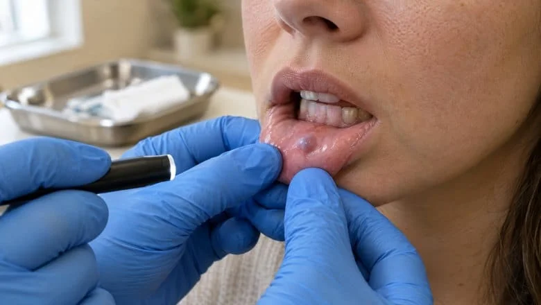
Lip lesion removal treats lumps, cysts, spots and patches on the lips: a mucocele (mucous cyst) inside the lip, a dark spot, a soft blue venous lake, a firm fibroma, or the rough sun damage of actinic cheilitis. Most lip lesions are harmless, but one of our plastic surgeons examines every one before treatment, and anything uncertain is sampled first.
Lip lesions are one part of all the skin lesions we remove, but the lip needs its own approach. The vermilion border, where the red lip meets the skin, shows even a small step, and the lips must still seal for speech and eating, so every excision is planned around both.
Surgical lip lesion removal and lip biopsy are carried out by consultant plastic surgeons, and laser treatment of a venous lake, a dark spot, a papilloma or actinic cheilitis by Ida Bafende, NMC-registered aesthetic practitioner, on the Fotona SP Dynamis Pro laser. Our guide to common skin lumps and bumps and our page on how skin lesions are assessed and removed cover the wider picture.
We remove benign lip lesions and treat pre-cancerous sun damage, but we do not treat skin cancer: a lip lesion that could be a skin cancer is referred through your GP to an NHS skin cancer team.
Lip lesion removal at a Glance
We see and treat adults aged 18 and over only. A new, changing or slow-healing lip lesion is biopsied or cut out for analysis rather than lasered, and your suitability for each technique is confirmed at your assessment.
Considering lip lesion removal?Book a consultation to have your lip lesion assessed and the right treatment explained. No obligation, and no pressure to book.
Lip lesion removal before and after photos
This photo shows a venous lake on the lip before and after laser treatment at Centre for Surgery, shared with the patient’s consent. A venous lake is a soft, dark blue or purple bump caused by a widened small vein, and it is usually treated with laser rather than surgery.
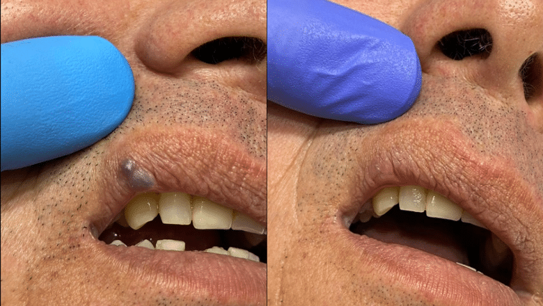
Individual results vary. A larger venous lake can need a second session, and a small pink mark can remain for a few weeks while the lip settles.
What is a lip lesion? Common lumps and spots on the lips
A lip lesion is any lump, spot, patch or sore on the lip. Where it sits is a strong clue to what it is:
- Lip skin: gets the same lesions as other facial skin, such as moles, cysts, seborrhoeic keratoses and, occasionally, a basal cell carcinoma.
- Vermilion border: the sharp line between the red lip and the skin, where a scar or step shows most.
- Vermilion (red lip): thin, sun-exposed tissue where actinic cheilitis, melanotic macules, venous lakes and lip squamous cell carcinoma usually appear, mostly on the lower lip.
- Inner lip lining: the moist mucosa, full of tiny salivary glands, where mucoceles, fibromas and papillomas form.
What are the most common lip lesions?
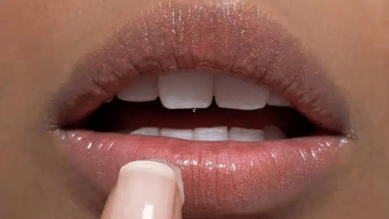
Most lip lesions are benign. The three we see most are mucoceles, venous lakes and labial melanotic macules, followed by fibromas, squamous papillomas and pyogenic granulomas. Fordyce spots, the tiny pale yellow dots many people have on their lips, are normal oil glands rather than a lesion.
A smaller group is pre-cancerous: actinic cheilitis and leukoplakia (a white patch on the lip lining that does not rub off), which can be biopsied to check the cells. Lip cancer is uncommon and is covered further down this page. If what bothers you is the size of your lips rather than a lump or spot, see lip reduction surgery.
Mucocele on the lip: what causes a lip cyst and how is it removed?
A mucocele (also spelt mucocoele, and often called a mucous cyst or lip cyst) is the most common lump inside the lower lip. It forms when the duct of a tiny minor salivary gland in the lip lining is damaged, usually by biting the lip, and saliva leaks into the tissue. The result is a soft, bluish or see-through dome, usually 5–15 mm across, that rarely hurts but can burst and refill, and a large one can get in the way of eating.

How to get rid of a mucocele
A small mucocele can settle on its own, so a new one is often watched for a few weeks. Piercing or draining it leaves the damaged gland behind, so it often refills.
A mucocele that persists or keeps coming back is removed as a day case under local anaesthetic, in 30–45 minutes. Through a small cut inside the lip, your surgeon removes the cyst with the gland feeding it and closes the lining with dissolving stitches. Marsupialisation, which opens the roof of the cyst so that it drains and heals, suits some mucoceles. The tissue is sent for histology.
Will a mucocele come back after removal?
Recurrence is uncommon when the feeding gland is removed, and more common when the cyst is only drained. The evidence is limited: in a small 2016 randomised comparison of 20 patients, 1 of 10 mucoceles came back after excision and 2 of 10 after micro-marsupialisation, a difference that was not statistically significant[1].
A cyst on the skin side of the lip line is more often an epidermoid cyst, treated with standard cyst removal.
Dark spot on the lip: labial melanotic macule or melanoma?
A flat brown or black spot on the lip, usually the lower lip, is most often a labial melanotic macule: a harmless patch of extra pigment, like a freckle on the lip, usually 3–10 mm across with a smooth edge, that stays the same for years. DermNet’s guide to the labial melanotic macule shows typical examples.
When is a dark spot on the lip a concern?
A lip melanoma is rare, but it can look similar at first. Have a pigmented spot checked if it is new in adult life, growing, uneven in shape or colour, raised, bleeding or changing. Our article on the difference between a mole and a melanoma explains the warning signs.
How is a dark spot on the lip checked?
One of our plastic surgeons examines it with a dermatoscope, a handheld magnifier with its own light. A 2018 retrospective comparative study in PLoS One (Lee and Mun) of lip lesions confirmed by histology found that venous lakes were mostly structureless and purple, red or blue under dermoscopy, while melanotic macules more often showed brown or grey lines and dots[2]. A spot that looks typical can be treated or left alone; one that does not is biopsied first.

Labial melanotic macule removal
A confirmed melanotic macule needs no treatment, but a prominent one can be removed by Ida Bafende with the Er:YAG laser mode of the Fotona SP Dynamis Pro, which removes the pigmented surface in thin layers, or as an excision biopsy, which also gives a sample for the laboratory. Laser leaves nothing to examine, so it is used only for a spot that is clearly benign at your assessment. We treat individual spots, not general darkening of the lips. A raised, mole-like spot at the edge of the lip is usually a mole on the lip.
Other lip lumps: venous lake, fibroma, papilloma and granuloma
Venous lake
A venous lake is a soft, dark blue or purple bump, usually 4–10 mm across, on the lower lip of an adult: a single widened small vein, mainly caused by years of sun exposure. It pales when pressed, can bleed if bitten or knocked, and has no cancer potential.
It is usually treated with the long-pulse Nd:YAG (1064 nm) mode of the Fotona SP Dynamis Pro, which heats the blood in the vessel and seals it. In a UK series of 31 consecutive patients with venous lesions of the lips and around the mouth, treated with a long-pulsed Nd:YAG laser, 27 had no sign of recurrence at an average follow-up of 12 months[3]. A larger venous lake can be excised instead; our venous lake removal page has the details.
Lip fibroma
A firm, smooth, skin-coloured lump inside the lip, caused by repeated rubbing or biting. It does not usually go away on its own and is removed by a simple excision, then sent for histology.
Squamous papilloma
A small white or pink growth with a bumpy, cauliflower-like surface. Some are linked to the human papillomavirus (HPV), which also causes warts. It is removed by excision or laser ablation.
Pyogenic granuloma
A bright red, fast-growing lump that bleeds easily, often after a minor injury. It is benign but troublesome, and on the lip it is excised and sent for histology; see pyogenic granuloma removal.
Other lip lumps and red spots
A soft, fatty lump inside the lip is occasionally a small mucosal lipoma, removed by excision. A tiny bright red dot on the lip skin is often a cherry angioma rather than a venous lake.
Actinic cheilitis treatment: sun damage on the lower lip
Actinic cheilitis is pre-cancerous sun damage to the lip, the lip form of the rough patches covered on our actinic keratosis treatment page. It almost always affects the lower lip, mostly in people with fair skin and years of outdoor work or sun exposure; smoking and alcohol add to the risk. DermNet’s page on actinic cheilitis has photographs.
Actinic cheilitis symptoms
- A lower lip that feels constantly dry, rough or chapped and does not improve with lip balm
- Scaling, cracking or flaking, sometimes with burning
- A blurred line where the red lip meets the skin
- White or grey patches, or a thickened, firm area
Why actinic cheilitis is treated
Actinic cheilitis can progress to squamous cell carcinoma, which on the lip is more likely to spread than the same cancer on other skin[4]. The risk for any one person is small but real. A 2019 systematic review and meta-analysis in Oral Diseases (Carvalho et al.) pooled 283 cases from 10 studies: remission was 92.8% after surgical treatment against 65.9% after non-surgical treatment, recurrence 8.4% against 19.2%, and about 2.5% of surgically treated cases showed malignant change[5]. The authors called for more standardised studies.
How is actinic cheilitis treated?
A thickened, ulcerated or firm area is biopsied first, to check it has not already become a cancer. Everyone needs a daily SPF 50+ lip balm. A small, well-defined patch can be excised, or treated by Ida Bafende with Er:YAG laser ablation, which removes the damaged surface in thin layers (see our article on the erbium laser). Freezing (cryotherapy) is widely used elsewhere for actinic cheilitis, but we do not offer it here; we use excision or the laser instead. For damage across much of the lip, some people are prescribed a cream by their GP or a dermatologist, and extensive damage needing a vermilionectomy (surgical removal of the surface of the red lip) is referred to a head and neck or reconstructive surgery team. Actinic cheilitis can recur after any treatment, so your clinician also agrees how your lip will be followed up.
What does lip cancer look like? Warning signs on the lip
Lip cancer usually starts on the lower lip as a sore, crust or thickened patch that does not heal. The NHS page on symptoms of mouth cancer includes a lump on the lip and a mouth ulcer lasting more than 3 weeks. Get checked if you notice:
- a sore, ulcer or scab on the lip that has not healed after 3 weeks
- a firm or thickened patch, a growing lump, or bleeding without an injury
- a new dark spot that is irregular in shape or colour, or changing
- a white or red patch on the lip lining that does not rub off
Types of lip cancer
Squamous cell carcinoma (SCC) is the most common: a crusted sore, scaly firm patch or ulcer on the lower lip, usually in older adults with heavy sun exposure or a smoking history, often where actinic cheilitis was. It can spread to the lymph glands in the neck: a 2024 systematic review and meta-analysis in Head & Neck (Kansara et al.) of 189 sentinel node biopsies found hidden spread in 21 (11.1%) and put the true rate at about 10%[6], which is why the neck is examined as part of lip SCC care.
Basal cell carcinoma (BCC) more often affects the skin of the upper lip, as a slow-growing pearly bump with tiny visible blood vessels that very rarely spreads. Melanoma of the lip is rare, showing as a dark, irregular, changing patch; a suspected lip melanoma is referred to an NHS skin cancer team rather than treated here.
What happens if a lip lesion could be cancer?
We remove benign lip lesions and treat pre-cancerous sun damage, but we do not treat skin cancer at Centre for Surgery. If a lip lesion looks as though it could be a skin cancer, or a biopsy shows one, we refer you through your GP to an NHS skin cancer team, on the urgent suspected cancer pathway where that applies, and we provide our consultation notes for that team. A biopsy here is only for a lip lesion whose nature is uncertain: one that clearly looks like a cancer is referred rather than cut out.
A confirmed lip cancer, even an early one, is referred to an NHS team for treatment and any reconstruction of the lip. Our guide to whether a lump needs removing lists the signs to look for.
How is a lip lesion diagnosed? Assessment and lip biopsy
Your consultation at our Baker Street hospital lasts about 30–45 minutes, longer if a biopsy is needed or several lesions are checked. One of our plastic surgeons asks how the lesion has changed and whether it bleeds, crusts or fails to heal, along with your sun exposure, smoking and general health, then examines the lesion, the rest of the lip, any pigmented spot with a dermatoscope, and the lymph glands in your neck where the type of lesion calls for it.
A clearly benign lesion can go straight to a treatment plan. Anything that does not look clearly benign at your assessment is not lasered: if its nature is uncertain, one of our plastic surgeons removes it surgically and sends it for analysis, or takes a small sample first, and a lesion that looks like a skin cancer is referred, as explained above.
We recommend a two-week cooling-off period between your consultation and treatment. If you have had a virtual consultation first and sent us photos of the lesion, treatment can take place at the same visit as your in-clinic assessment, as long as the surgeon (or Ida, for laser) is happy it is suitable to treat on the day.
Lip biopsy: when and how
A biopsy is recommended when the nature of a lip lesion is uncertain, for example if it is changing, growing, ulcerated, crusting, firm, slow to heal or irregularly pigmented. It is done under local anaesthetic, often at the same visit, and takes about 15 minutes. A punch biopsy removes a small 3–6 mm core, which suits a patch of actinic cheilitis; an excision biopsy removes the whole lesion, diagnosing and treating a small one in one step.
What happens to the tissue?
Tissue that is cut out (an excision, shave, punch or biopsy) is sent to an accredited laboratory for histology, where a pathologist examines it under the microscope. Laser destroys the tissue, so there is nothing to examine: that is why anything uncertain is cut out rather than lasered. Results usually take 3–4 weeks, and we contact you with the result.
Our guide to what happens to your histology sample explains the report, and our article on why removed tissue is sent for analysis sets out the reasoning.
How is a lip lesion removed? Excision, W-plasty and laser
The technique depends on what the lesion is, its size and depth, and where on the lip it sits. Techniques used elsewhere on the body are covered on our skin lesion assessment and removal page.
Excision with direct closure
Suits most small lip lesions. The lesion is cut out as a small ellipse and closed with fine stitches, lined up so that the vermilion border stays continuous, in 30–45 minutes under local anaesthetic.
W-plasty and Z-plasty
Used for slightly larger excisions, where a straight closure would pull the lip out of shape. A W-plasty breaks the scar into a zigzag of small triangles that is harder to see, which suits vertical scars on the lip; a Z-plasty swaps two small triangular flaps so that the scar does not shorten the lip or distort the vermilion border. Either takes 45–90 minutes under local anaesthetic.
Wedge excision
Used for a larger lesion of the red lip, up to about a third of its width. A V- or W-shaped full-thickness wedge is removed and the lip is closed in three layers (lining, muscle and skin), with the vermilion border aligned to avoid a step. It takes 60–90 minutes under local anaesthetic.
Laser treatment for lip lesions
Laser is used only for lesions that are clearly benign at assessment, because it leaves no tissue for the laboratory. Before any laser treatment, one of our plastic surgeons examines the lesion, with a dermatoscope where needed, to confirm it is benign and suitable for laser; Ida Bafende then carries out the laser treatment on the Fotona SP Dynamis Pro. She treats venous lakes with the long-pulse Nd:YAG (1064 nm) mode, which coagulates the vessel, with laser safety goggles or opaque shields over your closed eyes; everyone in the room wears eyewear rated for the laser. The Er:YAG (2940 nm) mode vaporises tissue in thin layers with little heat, and she uses it for a melanotic macule, a small papilloma or a small patch of actinic cheilitis.
Anaesthetic and sedation
Most lip procedures are done under local anaesthetic alone, and you stay awake. Sedation can be added for a larger excision or if you are anxious; our guide to local anaesthetic and sedation explains the options.
Will lip lesion removal leave a scar?
Any cut through the skin of the lip leaves a scar, so the aim is a fine, well-placed line. The most noticeable problem on the lip is a small notch or step in the vermilion border, so closures are lined up along that border and, where needed, broken up with a W-plasty or Z-plasty.
- After an excision on the lip skin or red lip: the scar is pink and slightly raised at first, and usually settles to a fine, pale line over 6–12 months.
- After mucocele removal: the cut is inside the lip, so the scar sits on the lining, where it is not usually seen.
- After laser for a venous lake: a small pink mark fades over 2–6 weeks. A scar is possible but uncommon: in one series, 1 of 31 patients had a small contracted scar[3].
Early scars look worse than they will become. Our guide to how facial scars fade explains the timeline, and a scar that is still raised or uneven after a year, or a firm lump of scar tissue from an old lip injury, can often be improved with scar revision.
Lip lesion removal side effects and risks
Lip lesion removal is minor surgery and serious complications are uncommon, but every procedure carries some risk, and your clinician explains the ones that apply to you.
- Swelling and bruising: expected, usually for 3–5 days after mucocele removal, 5–7 days after a simple excision and 7–10 days after a W-plasty, Z-plasty or wedge excision.
- Bleeding: light oozing in the first day is common, as the lip has a rich blood supply; firm pressure with clean gauze usually stops it.
- Infection: uncommon; signs are increasing pain after 48 hours, spreading redness, pus or a fever.
- Scarring: a visible line, a small notch in the vermilion border, or a raised scar.
- Altered feeling: numbness or tingling around the scar, which usually improves over the following months.
- Recurrence: a mucocele can return if a feeding gland is left behind, and a venous lake can need a second laser session.
- Colour change after laser: the area can heal paler or darker than the rest of the lip.
- Further treatment: occasionally histology shows a lesion was not what it appeared to be, or was not fully removed.
Smoking slows lip healing and raises the risk of a poor scar, so we advise stopping 4 weeks before and 4 weeks after surgery.
Lip lesion removal recovery and aftercare
After laser treatment of a venous lake
The lip is red and slightly swollen at first, and a small dark crust forms within 24 hours, then lifts over 7–14 days. Most people go back to work the same day. The food, drink, alcohol and sun advice below applies after laser too.
After mucocele removal
The inside of the lip is swollen and tender for 3–5 days, and the stitches dissolve over 7–10 days. Eat soft food for 3–5 days, avoid acidic and spicy food for a week, and rinse with warm salt water 3 times a day. Most people return to work within 1–2 days.
After an excision, W-plasty, Z-plasty or wedge excision
Expect swelling and tightness for 5–7 days after a simple excision and 7–10 days after a reconstruction. Skin stitches come out at 5–7 days (at 7 days after a W-plasty or Z-plasty); stitches inside the lip dissolve. Eat soft food for 3–5 days (5–7 after a reconstruction), avoid opening your mouth wide for 7–14 days, and sleep with your head slightly raised for the first few nights. Most people return to work within 1–3 days of a small excision and 3–5 days of a reconstruction; a larger excision can need 5–7 days off.
Aftercare for every lip procedure
- Keep a wound on the outside of the lip moist with petroleum jelly twice a day until it has healed.
- Avoid alcohol for 48 hours, very hot food and drink for 24–48 hours while the lip may be numb, and spicy or acidic food for a week.
- Avoid contact sports, or anything that risks a knock to the lip, for 2 weeks.
- Keep direct sun off the lip for at least 6 weeks, then use an SPF 50+ lip balm every day.
- For pain, paracetamol is usually enough; avoid ibuprofen for the first 48 hours. If you take aspirin that was not prescribed, stop it 2 weeks before your procedure; if your doctor prescribed it, ask them first.
Scar care once healed
Once the wound has healed, usually at about 2 weeks, use SPF 50+ lip balm daily to stop the scar darkening, and from week 4 to week 12 use gentle massage and a silicone gel or sheet (bought separately) to soften it. After a sun-related lesion such as actinic cheilitis, daily lip sun protection is the main way to prevent new damage.
When to contact us
Call us on 020 7993 4849 if you have increasing pain after 48 hours, redness spreading beyond the wound, pus, a fever, a wound that opens, or bleeding that does not stop with firm pressure.
How much does lip lesion removal cost in London?
The cost of lip lesion removal depends on what the lesion is, the technique, its size and whether the lip needs reconstruction, how many lesions are treated, and whether sedation is added. Our lip lesion removal prices are listed in the Skin & Aesthetics price list, which has separate options for:
- mucocele excision
- laser treatment of a venous lake, or its excision
- removal of a labial melanotic macule
- removal of another small benign lip lesion
- pyogenic granuloma excision
- a punch biopsy
- wedge excision, and excision with W-plasty or Z-plasty
- a course of actinic cheilitis treatment
- sedation, if needed
Your price is confirmed after your assessment, once the lesion has been examined and the technique agreed. The consultation fee is redeemable against your treatment. Where tissue is sent for analysis, the laboratory invoices you directly.
Finance is available through Chrysalis Finance, subject to status; see our finance options page. M Sarwar Limited, trading as Centre for Surgery, is a credit broker, not a lender.
Why choose Centre for Surgery for lip lesion removal
Surgical lip lesion removal here is carried out by a GMC-registered consultant plastic surgeon, who plans each excision around the vermilion border and the way your lips move. Laser treatment of venous lakes, dark spots, papillomas and actinic cheilitis is carried out by Ida Bafende, NMC-registered aesthetic practitioner, on the Fotona SP Dynamis Pro, after one of our plastic surgeons has examined the lesion. Having biopsy, excision, lip reconstruction and laser in one place means the method can be chosen to suit your lesion.
Centre for Surgery is rated Good overall by the CQC. You get honest advice, including when a lip lesion is better left alone or when a referral is the right next step, and there is no pressure to book. You can read our patient reviews before you decide.
Consultations and procedures take place at our hospital at 95–97 Baker Street. To book, use the consultation form on this page or call 020 7993 4849.
Lip lesion removal research and references
The studies cited on this page:
- Giraddi GB, Saifi AM. Micro-marsupialization versus surgical excision for the treatment of mucoceles. Annals of Maxillofacial Surgery. 2016;6(2):204-209. doi:10.4103/2231-0746.200324
- Lee JS, Mun JH. Dermoscopy of venous lake on the lips: a comparative study with labial melanotic macule. PLoS One. 2018;13(10):e0206768. doi:10.1371/journal.pone.0206768
- John HE, Phen HS, Mahaffey PJ. Treatment of venous lesions of the lips and perioral area with a long-pulsed Nd:YAG laser. British Journal of Oral and Maxillofacial Surgery. 2016;54(4):376-378. doi:10.1016/j.bjoms.2015.06.014
- Trager MH, Farmer K, Ulrich C, et al. Actinic cheilitis: a systematic review of treatment options. Journal of the European Academy of Dermatology and Venereology. 2021;35(4):815-823. doi:10.1111/jdv.16995
- Carvalho MV, de Moraes SLD, Lemos CAA, et al. Surgical versus non-surgical treatment of actinic cheilitis: a systematic review and meta-analysis. Oral Diseases. 2019;25(4):972-981. doi:10.1111/odi.12916
- Kansara S, Oral E, Sarkar I, et al. Rate of occult metastasis in lip squamous cell carcinoma: a systematic review and meta-analysis. Head & Neck. 2024;46(10):2517-2523. doi:10.1002/hed.27747
What To Expect
Your lip lesion consultation
Your assessment takes about 30–45 minutes at our Baker Street hospital. One of our plastic surgeons examines the lesion, uses a dermatoscope on any pigmented spot, and explains whether a biopsy is needed first, which technique suits it and the price.
We recommend a two-week cooling-off period before treatment. If you had a virtual consultation and sent us photos first, treatment can take place at this visit when your lesion is suitable to treat on the day.
On the day of your procedure
The lip is numbed with a local anaesthetic injection, or with numbing cream before laser. A biopsy takes about 15 minutes, most excisions 30–45 minutes, and a wedge excision or W-plasty up to 90 minutes.
Any tissue that is cut out goes to the laboratory. You go home the same day with written aftercare advice and a number to call if you are worried.
After your lip lesion removal
Expect swelling and tenderness for a few days, longer after a reconstruction. Eat soft food, avoid alcohol for 48 hours, very hot food and drink for 24–48 hours and spicy or acidic food for a week, and keep an outer lip wound moist with petroleum jelly.
Skin stitches come out at 5–7 days, and stitches inside the lip dissolve. Most people are back at work within 1–3 days, or the same day after laser.
Your histology result and final result
If tissue was sent to the laboratory, results usually take 3–4 weeks, and we contact you with the result and explain what it means, including any next step.
A laser crust lifts within 7–14 days. A surgical scar fades from pink to pale over 6–12 months; protect it with SPF 50+ lip balm every day. A scar inside the lip, after mucocele removal, is not usually seen.
FAQs
-
Most lip lesions have a simple cause. Biting or rubbing the lip causes mucoceles and fibromas; years of sun exposure cause actinic cheilitis and venous lakes and raise the risk of lip cancer; extra pigment causes labial melanotic macules; the human papillomavirus is linked to some papillomas; and a minor injury can set off a pyogenic granuloma. Cold sores, caused by the herpes simplex virus, are a very common cause of a sore on the lip, but they heal on their own and are not removed.
-
Sometimes, yes. A small mucocele can burst and heal within a few weeks, especially if you stop biting or sucking the area. If one is still there after a few weeks, keeps refilling, grows, or gets in the way of eating or speaking, it is less likely to settle, because the damaged salivary gland keeps leaking. Removing the cyst together with its feeding gland is then the usual treatment. A lump that is firm, bleeds or does not behave like a mucocele should be checked.
-
No. Piercing or squeezing a mucocele lets the saliva out for a while, but the damaged gland underneath keeps leaking, so it often fills up again, and breaking the lining of the mouth risks infection and scarring. Home remedies cannot remove the gland either. If a mucocele has not settled after a few weeks, it can be removed with its gland under local anaesthetic, through a small cut inside the lip.
-
You feel the local anaesthetic injection, a sharp sting for a few seconds, and after that the lip is numb, so the removal itself should not hurt, although you may feel pressure or movement. Once the anaesthetic wears off, the inside of the lip is usually sore and swollen for 3–5 days. Paracetamol is generally enough, along with soft food and warm salt-water rinses. If you are anxious about the procedure, sedation can be added.
-
A true cyst, such as an epidermoid cyst in the skin, has its own lining wall and fills with keratin. Most mucoceles have no true lining: they are a pool of saliva that has leaked from a damaged salivary duct into the tissue of the lip. A mucocele sits inside the lip and looks bluish and see-through, while a skin cyst sits under the skin of the lip line. Both are removed surgically, and skin cysts are covered on our cyst removal page.
-
Usually not. Most single dark spots on the lip are labial melanotic macules, which are harmless flat patches of pigment, or venous lakes, which are small blue-purple blood vessels. Melanoma of the lip is rare. Have a dark spot checked if it is new in adult life, growing, irregular in shape or colour, raised, bleeding or changing; one of our plastic surgeons examines it with dermoscopy and, if there is any doubt, takes a biopsy. Our article on why removed tissue is checked explains what the laboratory looks for.
-
A cold sore starts with tingling, then a cluster of small blisters that crust over and heal within a week or two. Several other lip lesions can look similar at first: a mucocele, a pyogenic granuloma, impetigo, the cracked, scaly patches of actinic cheilitis and, rarely, an early lip cancer. The difference is how they behave. A sore that does not heal, keeps returning to the same spot without blistering, or bleeds easily is not acting like a cold sore. The NHS lists a lump on the lip and a mouth ulcer lasting more than 3 weeks among the symptoms to have checked by a GP or dentist.
-
No. A venous lake is a widened small vein near the surface of the lip, common in older adults after years of sun exposure, and it has no cancer potential. It can bleed if you bite or knock it, and some people dislike how it looks. The main reason to have it checked is that a dark lip spot can occasionally be something else, such as a melanoma, so one of our plastic surgeons examines it with dermoscopy first. Our venous lake treatment page explains removal.
-
No. Actinic cheilitis is not cancer, but it is pre-cancerous: years of sun damage have changed the cells of the lower lip, and a small proportion of cases go on to become squamous cell carcinoma. A patch can already have changed by the time it is noticed, so any thickened, ulcerated or firm area is biopsied before treatment. It is the lip form of actinic keratosis, and the same principles apply as for actinic keratosis on the face.
-
Lip lesion removal is minor surgery under local anaesthetic, and serious complications are uncommon. The usual effects are swelling, bruising and tenderness for a few days. Less common risks include infection, bleeding, a visible scar or a small notch in the line of the lip, numbness around the scar, and the lesion coming back. The risk of a noticeable notch is the reason closures are planned along the vermilion border. Your surgeon explains the risks that apply to your lesion before you agree to treatment.
-
It depends on the lesion. A mucocele can return if a feeding salivary gland is left behind, which is why the gland is removed with it. A venous lake treated with laser occasionally needs a second session. Actinic cheilitis can recur, and new patches can form on sun-damaged lips, so daily SPF 50+ lip balm matters in the long term. Most other benign lesions are unlikely to come back once fully removed, but new ones can form. If histology shows a lesion was not fully removed, your surgeon explains whether more treatment is needed.
-
The price depends on the size and position of the mucocele and whether sedation is added, and it is confirmed after your assessment. Mucocele excision has its own option in our lip lesion removal prices, alongside the other lip treatments. Where tissue is sent for analysis, the laboratory invoices you directly.
-
Sometimes. Your GP or dentist can examine a lip lesion, and one that could be cancer can be referred on the urgent suspected cancer pathway. Removal of a harmless lesion for cosmetic reasons is not normally available on the NHS, and whether a troublesome benign lump, such as a mucocele that keeps refilling, is treated depends on your GP’s assessment and local policy. Many people choose private lumps and bumps removal to have a benign lesion checked and removed at a time that suits them.


