

Real otoplasty and ear surgery results from patients treated at our Baker Street day surgery facility. Jump to Otoplasty & Ear Pinning, Ear Reshaping & Symmetry, Earlobe Repair or Earlobe Reduction.
Results vary from patient to patient. All photos are published with written patient consent and are examples of outcomes achieved at Centre for Surgery, not guaranteed results. After photos were taken at least six months after surgery unless the caption says otherwise. No photograph has been retouched or filtered; some are cropped or masked only to protect patient privacy. Full disclaimer below, and how to read these photos — including why the rear view is the only one that tells you the truth.
Ear pinning before and after — otoplasty for prominent ears
4 photographs
Otoplasty, ear pinning and pinnaplasty all describe the same procedure: setting back ears that project further from the head than average. Read more about what ear pinning involves, where the scars sit, and whether ears can go back after surgery.
Otoplasty — also called ear pinning or pinnaplasty — reduces the projection of prominent ears by reshaping the cartilage and repositioning the ear closer to the head. The aim is a natural ear-to-head angle rather than an over-corrected, flattened appearance. Learn more on our otoplasty surgery page.


Ear Reshaping and Symmetry Correction
7 photographs
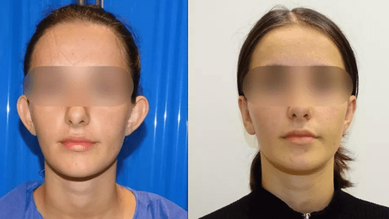
Not every otoplasty is about protrusion. Ear reshaping corrects asymmetry, irregular cartilage folds and differences in ear size or shape between the two sides. A well-executed result blends harmoniously with the rest of the facial features rather than drawing attention to the ears.


Earlobe Repair Before and After
14 photographs
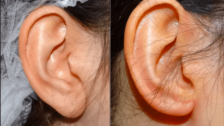
Earlobe repair restores a split, torn or stretched earlobe using a small excision and layered closure, usually under local anaesthetic. The cases below cover complete splits, enlarged piercing holes, and stretched or gauged earlobes. Full details on our earlobe repair page.
Split Earlobe Repair


Incomplete Split and Enlarged Piercing Hole Repair


Stretched and Tribal Earlobe Repair


Earlobe Reduction Before and After
6 photographs
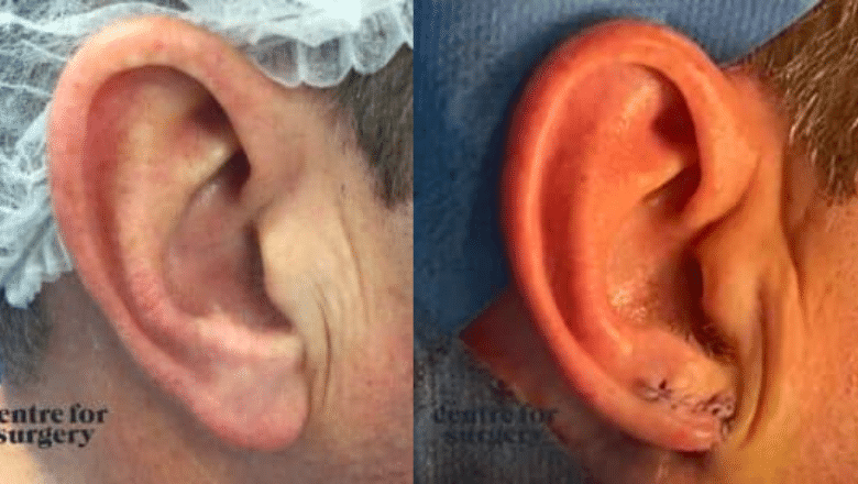
Earlobe reduction shortens elongated or pendulous earlobes, whether caused by ageing, long-term heavy earring use, or simply genetics. A crescent of tissue is removed from the inferior lobe margin and the incision is hidden within the natural contour. See our earlobe reduction page for full details.




About These Otoplasty Before and After Photos
The otoplasty before and after photos above are presented for educational purposes only and should be interpreted subjectively. They do not represent the opinion or view of any patient regarding their result, and they are not intended to suggest the result that any other patient may achieve. No two patients are alike, and no two results are necessarily alike. Only a personal consultation with your surgeon can provide you with a reasonable expectation of the results you may achieve from any specific procedure. We only publish photographs of patients who have given full written consent for their images to be used.
How to Read Otoplasty Before and After Photos
An ear is a three-dimensional structure, and a photograph flattens it. Almost every mistake an otoplasty can make is invisible from the front — which is the view most galleries publish. Knowing which view to demand, and what to look for in it, is worth more than looking at a hundred results.
Insist on the rear view
Ear prominence is a distance: how far the rim of the ear stands away from the side of the head. Only a photograph taken from behind lets you see that distance on both ears at once and compare them. It is the view in which asymmetry becomes obvious, and it is therefore the view most often left out. A gallery of front-facing photographs is showing you the angle in which prominent ears are least visible and corrected ears are hardest to judge. Ours includes rear views for exactly that reason.
Look for the telephone deformity
The ear should be brought back along its whole length. If the middle of the ear is over-corrected while the upper pole and the earlobe are left projecting, the silhouette comes to resemble an old telephone handset — narrow in the middle, flared at both ends. The reverse pattern, where the middle is left proud, is also possible. Both are visible in a profile or rear photograph and neither can be seen from the front.
From the front, the rim should still be visible
In a natural ear viewed head-on, the outer rim remains just visible beyond the fold of cartilage inside it. Over-pinning hides the rim behind the fold and produces the flattened, plastered-back appearance that reads immediately as surgical. Less correction, done evenly, looks more natural than more correction.
Look at the fold, not just the position
Otoplasty works by creating or reinforcing the natural fold of cartilage in the ear. Done well, that fold is a soft, continuous curve. Tightened too aggressively, it becomes a sharp ridge or a visible crease — an ear in the right place with the wrong shape. Close-up photographs show this; distant ones do not.
Check the head angle and the hair
A small rotation of the head changes how far an ear appears to project. So does hair tucked behind one ear and not the other. In an honest pair, the head is square to the camera in both frames and the hair is treated the same way in each.
For earlobe photographs, check the scale
Earlobe repair and reduction results are judged on a scar a few millimetres long. A closer crop, a softer light or a slightly different angle can make that scar disappear. Ask at what interval the photograph was taken — a fresh scar and a mature one look nothing alike.
What Stage of Healing You’re Looking At
Cartilage has memory, and this makes otoplasty photographs unusually sensitive to timing.
In the first weeks the ears are swollen and often bruised, and a supportive headband is worn as directed by your surgeon. That much is obvious in any early photograph. What is less obvious is what happens afterwards: cartilage that has been reshaped and held with sutures tends, to some degree, to want to return towards its original position. Most of any such change occurs over the first months. This means an ear photographed shortly after surgery may sit slightly closer to the head than the same ear photographed a year later, and a small proportion of patients seek revision for partial recurrence of prominence.
The consequence is the opposite of a facelift, where the early photograph flatters through swelling. Here the early photograph flatters through cartilage that has not yet finished settling. A gallery that never states the interval is asking you to assume the most favourable point on that curve.
Scars sit behind the ear, in the crease where it meets the head. They pass through a firm, pink phase before softening, and in this location thickened or keloid scarring can develop months after an otherwise uneventful recovery — something no photograph taken at six weeks can rule out. Earlobe scars follow a similar course, typically fading substantially over three to six months, and re-piercing is generally deferred for at least six months after a repair.
The after photographs in this gallery were taken at least six months after surgery, unless the caption says otherwise. For what to expect at each stage before then, see our otoplasty recovery guide.
What These Photographs Cannot Show You
There is a limit to what any gallery can tell you, and a clinic that pretends otherwise is selling rather than informing.
- Whether the ears stayed there. Reshaped cartilage can partially relapse, and when it does it does so over months. Every photograph here was taken at a single moment. The honest question is how the ears look at two years, and only your surgeon’s own follow-up can answer it.
- The scar. It sits behind the ear, in the one place a front, profile or rear photograph of a corrected ear does not look. Its absence from these images is not evidence of its absence from the patient.
- How the ear feels. Numbness, tenderness to pressure, and sensitivity to cold are common for a period after surgery. An ear that has been folded too sharply can also be felt as a ridge by the person who owns it, long after it stops being visible to anyone else.
- True symmetry. Ears are rarely identical before surgery. They differ in size, in shape, and in where they sit on the skull — one is often set higher than the other. A photograph flattens all three. Surgery corrects projection; it cannot make two different ears the same ear, and a result that respects a patient’s existing asymmetry is a better result than one that pretends to erase it.
- Whether surgery was the answer. Prominent ears are a normal anatomical variation, not a deformity. Where the patient is a child, what matters is how the child feels rather than how the ears photograph, and that is a conversation rather than a comparison.
None of this is a reason to distrust before and after photographs. It is a reason to treat them as the beginning of a conversation with a surgeon rather than the end of one.
Otoplasty Results: Common Questions
Is otoplasty a big operation?
No. Otoplasty is usually a day case lasting one to two hours, often performed under local anaesthetic, with the incisions hidden behind the ears. Recovery is mainly about protecting the ears: a headband is worn, particularly at night, for several weeks while the cartilage sets in its new position.
Is there a non-surgical option for ear pinning?
For adults, no non-surgical treatment permanently changes the shape of the ear cartilage. Splinting can work in newborn babies, whose cartilage is still soft, but not later in life. Stitch-only techniques use smaller incisions but suit a limited range of ears and are more likely to let the ears move back.
Is otoplasty risky?
Serious complications are uncommon. Recognised risks include bleeding, infection, the ears moving back towards their original position, asymmetry, problems with the stitches, thickened scars behind the ear and over-correction. Your surgeon will discuss which of these apply to you before you decide.
Otoplasty and Ear Surgery at Centre for Surgery
Centre for Surgery is a private hospital for plastic and cosmetic surgery on Baker Street in central London, registered with and independently inspected by the Care Quality Commission. Our surgeons perform otoplasty, ear pinning, ear reshaping, earlobe repair and earlobe reduction at our purpose-built day surgery facility.
Experienced Surgeons
Every surgeon on our team is GMC-registered and independently verifiable on the General Medical Council register. Ear surgery is performed by surgeons with specific experience in otoplasty and earlobe reconstruction.
Our Facility
Centre for Surgery operates from a purpose-built day surgery facility at 95–97 Baker Street, registered with the Care Quality Commission. Most ear procedures, including earlobe repair and reduction, are performed under local anaesthetic as a walk-in, walk-out procedure.
Personalised Approach
Our clinic emphasises the importance of a personalised approach to each patient, tailoring the otoplasty surgery to address the individual’s unique needs and goals. This ensures that the patient’s expectations are met and the results are natural-looking and harmonious.
Aftercare and Support
From post-operative follow-up appointments to being available to address any concerns or questions, the Centre for Surgery team supports you throughout your otoplasty journey. Our full scar management programme is included as standard.
Verified Patient Reviews
We invite you to read what our otoplasty patients say on independent, verified review platforms. Every review is publicly verifiable, dated, and linked to a real named patient.
- Google Reviews: Read our Google reviews — rated 4.6 stars based on hundreds of verified patient reviews.
- Patient reviews page: Read patient reviews by procedure — Google and Trustpilot reviews brought together on our own site.
As a CQC-regulated provider, every surgeon on our team is GMC-registered and independently verifiable. View our CQC rating.
Related Reading
- Will My Ears Look Natural After Otoplasty?
- Is Otoplasty Painful?
- How Long Should You Wear a Headband After Otoplasty?
Explore More Before and After Galleries
View our full collection of plastic surgery before and after photos, or browse related facial procedure galleries: Rhinoplasty Before and After Photos | Blepharoplasty Before and After Photos | Facelift Before and After Photos
Subject to status, we offer finance options with Chrysalis Finance. For a full price breakdown, see our otoplasty cost guide. Learn more on our About Us page, browse our Plastic Surgery Blog, or find answers in our Clinic FAQs.
To book a face-to-face consultation with a specialist otoplasty surgeon, call us today on 020 7993 4849 or complete the contact form below.