

In this guide10 sections
- Summary
- What does yours look like? A quick guide
- The most common types of eyelid lump
- How eyelid lumps are assessed
- Why specialist eyelid surgery matters
- How treatment is performed
- Combining lump removal with blepharoplasty
- When eyelid lumps warrant urgent assessment
- What we don’t recommend
- Frequently asked questions
Summary
- Most lumps on the eyelid are harmless. Chalazion, stye, milia, xanthelasma, syringoma and skin tags together account for the great majority.
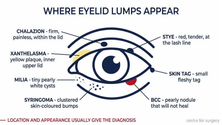
- Location and appearance usually give the diagnosis. A firm painless lump inside the lid is a chalazion; a red tender one at the lash line is a stye; a yellow patch at the inner upper lid is xanthelasma; a small white spot on the eyelid is usually a milium.
- A chalazion is not infected. It is a blocked oil gland, and squeezing it makes things worse. Warm compresses for four to eight weeks resolve most of them.
- The eyelid is a common site for basal cell carcinoma, particularly the lower lid. Any lesion that grows, bleeds, crusts or fails to heal needs assessing rather than watching.
- Xanthelasma is worth a cholesterol check. It is associated with raised lipids in a substantial proportion of people.
- Eyelid lesions should not be removed at a beauty clinic. The lid margin, lash line and lid closure all have to be preserved, and pathology can be missed.
Lumps on the eyelid are extremely common, and most are harmless. A bump on the eyelid can be one of around a dozen different things, and eyelid bumps are among the commonest reasons people ask a plastic surgeon to look at their eyes. The eyelid skin is thin, mobile, vascular and rich in specialised structures — oil glands, sweat glands, hair follicles, lymphatic channels — and lesions can arise from any of them. The vast majority are entirely benign. A smaller but important minority are clinically significant and warrant prompt assessment, particularly when they appear in older patients or fail to resolve over time.
This guide covers the most common types of eyelid lump, how they are distinguished from each other, when professional review is indicated, and what treatment looks like at Centre for Surgery’s CQC-regulated Baker Street private hospital. Eyelid surgery is one of the more technically demanding areas of plastic surgical practice — the anatomy is delicate, scars must be exceptionally fine, and the functional integrity of the eyelid must be preserved. This is not an area for non-specialist removal.
What does yours look like? A quick guide
Most people arrive here having noticed something specific — a white spot on the eyelid, a yellow patch, or a small lump that will not go away — without knowing what it is called. Colour, position and whether it hurts will usually narrow it down. Find the description that matches yours, then read the relevant section below.
| What you can see | How it feels | Most likely |
|---|---|---|
| A small white spot on the eyelid, or a white dot just under the skin surface | Firm, painless, pearly, doesn’t move | Milia |
| A yellow patch on the eyelid — often at the inner corner of the upper lid, sometimes both eyes | Flat or slightly raised, soft, painless | Xanthelasma |
| A hard painless lump in the eyelid, more visible when you pull the lid | Firm, well-defined, not tender once established | Chalazion |
| A red painful lump at the lash line, sometimes with a yellow head | Hot, tender, swollen | Stye |
| Several small flesh-coloured bumps clustered under the eyes | Soft, painless, symmetrical on both sides | Syringomas |
| A small dangling tag of skin on the lid | Soft, mobile, painless | Skin tag |
| A pearly or translucent nodule, possibly with tiny visible blood vessels, that bleeds, crusts or won’t heal | Firm, may ulcerate, slowly growing | Needs prompt assessment — possible BCC |
This is a guide to help you understand what you’re looking at — not a diagnosis. Anything persistent, growing, bleeding or changing should be examined in person, whatever colour it is.
The most common types of eyelid lump
Chalazion (meibomian cyst) — a hard, painless lump on the eyelid
A chalazion is the single most common eyelid lump. It forms when a meibomian gland — one of the oil-producing glands along the lid margin — becomes blocked, causing oily material to accumulate within the gland and trigger a local granulomatous inflammatory response. The result is a firm, painless, well-defined lump sitting within the eyelid, most commonly in the upper lid. If you have a hard lump on the eyelid that doesn’t hurt, this is the usual explanation.
A chalazion is not infected — it is an inflammatory rather than an infective lesion. It is not painful in its established form, though it may be uncomfortable in its early days when it first develops. Many chalazia resolve spontaneously over a few weeks with warm compresses, gentle lid massage, and good eyelid hygiene. Persistent chalazia — those that remain after 4–8 weeks of conservative management — are typically treated by surgical incision and curettage, performed under local anaesthetic from the inside of the eyelid (no visible external scar).
Stye (hordeolum) — a red, painful lump on the eyelid
A stye is an acute infection of a hair follicle or associated gland at the eyelid margin. Unlike a chalazion, it is genuinely infected — the lesion is red, hot, swollen and tender, with a yellow head of pus often visible. Most styes resolve within a week or two with warm compresses and good lid hygiene. Topical antibiotics are sometimes prescribed; surgical drainage is rarely needed.
An unresolved stye can occasionally evolve into a chalazion as the acute infection settles but the underlying gland blockage persists.
If you get chalazia or styes repeatedly, blepharitis is the usual reason. Blepharitis is chronic low-grade inflammation of the lid margins, and it keeps the meibomian glands congested. Treating each lump as it appears does not stop the next one; daily lid hygiene — warm compress, gentle lid-margin cleaning, and treating any associated rosacea or dry eye — reduces recurrence far more effectively. Anyone presenting with a third or fourth chalazion should be assessed for blepharitis rather than simply having another one drained.
Xanthelasma — yellow patches on the eyelid
If you have noticed something yellow inside the eyelid area or a yellow spot on the eyelid skin, xanthelasma is the most likely cause. Xanthelasma are yellowish, lipid-rich plaques that develop on the eyelid skin — most commonly on the upper inner eyelid, and often symmetrically on both eyes. They are not painful, do not affect vision, and are not dangerous. They are, however, frequently associated with elevated cholesterol levels — and patients with xanthelasma should generally have lipid screening as part of the assessment.
Worth distinguishing: a yellow appearance inside the lid, on the conjunctiva rather than the skin, is a different thing — that can be a pinguecula, or in the context of generally yellow eyes, a sign of jaundice, which needs medical review rather than cosmetic treatment. Xanthelasma sits on the eyelid skin, not the inner surface.
Treatment options for xanthelasma include erbium laser ablation (scarless surface treatment for most lesions), surgical excision (for larger or deeper lesions), trichloroacetic acid application, and radiofrequency ablation. Erbium laser is our preferred first-line technique for most xanthelasma. See also our guide to whether you can have xanthelasma with normal cholesterol.
Milia — small white spots on the eyelid
A white spot on the eyelid, or a tiny white dot that looks like it sits just beneath the skin surface, is most often a milium. Milia are small, pearly-white cysts that develop superficially in the skin, often around the eyes. They are filled with keratin and are entirely benign. They are firm rather than fluid-filled, which is why squeezing them does nothing except irritate the skin.
Again, position matters: a white dot on the inner surface of the lid or on the white of the eye is not a milium and needs a different assessment. Milia sit in the eyelid skin. Milia removal involves making a tiny incision in the overlying skin and extracting the contents — a quick, scarless procedure. Our guide to milia around the eyes covers why they recur and how to reduce that.
Skin tags
Small fleshy skin tags can develop on the upper or lower eyelid skin. They are entirely benign and easily removed under local anaesthetic with fine scissors or radiofrequency cautery. Skin tag removal on the eyelid requires specialist technique to avoid affecting the lid margin or lashes.
Syringomas
Syringomas are small, soft, flesh-coloured bumps that develop in clusters around the eyes, most commonly on the lower eyelids and upper cheek. They are benign growths of the eccrine sweat duct cells. If you have several small lumps on the eyelid rather than one, appearing symmetrically on both sides, syringomas are a likely explanation. Treatment is most often with fine ablative laser or radiofrequency, working through each individual lesion. Multiple sessions are typical.
Eyelid cysts
An eyelid cyst is most often a chalazion, but true epidermoid and pilar cysts can also develop on the eyelid, though less commonly than on the face proper. They present as smooth, mobile lumps and are managed by surgical excision with complete capsule removal — see cyst removal and our guide to sebaceous cyst vs epidermoid cyst.
Moles and pigmented lesions
Moles can develop on the eyelid skin or — less commonly — on the lid margin. Eyelid moles require specialist technique for removal to preserve the lid margin contour and function. Every excised mole at Centre for Surgery is sent for histological analysis as standard. See mole removal.
Basal cell carcinoma and other skin cancers
The eyelid is one of the most common sites for basal cell carcinoma (BCC), the most common form of skin cancer. BCCs typically appear as pearly, translucent or skin-coloured nodules with small blood vessels visible on the surface; they may ulcerate or fail to heal. Lower eyelid BCC is particularly common because of cumulative UV exposure to this area over a lifetime.
Any persistent, ulcerating, or unusual eyelid lesion in an older patient warrants prompt assessment. Eyelid BCC requires specialist excision with histological margin control, often using Mohs micrographic surgery or formal histological assessment. Reconstruction of the eyelid after BCC excision is a specialist plastic surgical procedure.
How eyelid lumps are assessed
Assessment at Centre for Surgery typically takes 20–30 minutes and includes:
- History — how long the lesion has been present, whether it has changed, any associated symptoms (pain, vision changes, discharge), and your general medical and skin history.
- Visual examination — assessment of the lesion’s appearance, location, surface characteristics, and relationship to surrounding eyelid structures.
- Eyelid eversion if indicated — turning the eyelid inside out to inspect the inner surface, where chalazia and some other lesions are best visualised.
- Dermoscopy — for pigmented lesions or lesions where surface features are difficult to assess by naked eye.
- Discussion — explanation of what the assessment has shown, treatment options, expected cosmetic outcome, and quotation.
For benign-appearing lesions where the patient wishes to proceed, treatment can often be carried out in the same session. For lesions where histology is needed, surgical excision is arranged with formal pathological analysis.
Why specialist eyelid surgery matters
The eyelid is one of the most anatomically and functionally demanding areas in the body for surgical work. Several specific issues:
- Thin skin — eyelid skin is the thinnest on the body. It heals quickly, scars beautifully when handled well, but heals badly when handled poorly.
- Functional importance — the eyelid must close completely to protect the cornea. Poorly planned eyelid surgery can disturb closure, causing dry eye, exposure keratopathy or even corneal damage.
- Cosmetic visibility — eyelid scars are in plain view. Imprecise technique produces scars that are immediately noticeable.
- Lid margin sensitivity — any work near the lid margin must preserve the position of the lashes, the contour of the lid edge, and the function of the meibomian glands.
- Anatomical layers — the eyelid has multiple distinct anatomical layers (skin, muscle, fibrous tarsal plate, conjunctiva), and surgical technique must respect each.
- Proximity to vision — work close to the visual axis demands precision, gentle tissue handling and complete absence of cautery damage near the cornea.
For these reasons, eyelid surgery — including the surgical removal of lumps and bumps — should be performed by a surgeon with specific expertise in eyelid surgery. At Centre for Surgery, surgical eyelid procedures are performed by GMC-registered plastic surgeons with extensive blepharoplasty experience, and lesions on the eyelid margin itself are removed by one of our facial plastic surgeons. Laser treatment of eyelid lesions such as milia, skin tags, syringomas and xanthelasma is carried out by Ida Bafende, our NMC-registered aesthetic practitioner, after one of the plastic surgeons has examined the lesion.
How treatment is performed
Most eyelid lump removals at Centre for Surgery are performed under local anaesthetic as day-case procedures. The patient remains awake throughout, the eyelid is fully numbed before any incision is made, and most patients leave the clinic within an hour of arrival. Specific techniques:
- Chalazion — incision and curettage from the inside (conjunctival) surface of the eyelid, leaving no visible external scar. 10–15 minute procedure.
- Xanthelasma — erbium laser ablation in most cases (scarless), surgical excision for larger or deeper lesions.
- Skin tags and small lesions — fine scissor excision or radiofrequency removal.
- Milia — small incision in the overlying skin and extraction.
- Syringomas — fine ablative laser or radiofrequency, treating each lesion in turn.
- Cysts — surgical excision with complete capsule removal, with skin closure using fine sutures.
- Moles — surgical excision with histological analysis as standard. Closure technique calibrated to the specific location.
- Suspicious lesions — not treated at Centre for Surgery, which does not treat skin cancer. A lesion that looks like a skin cancer is referred through your GP to an NHS skin cancer team.
For broader context on what eyelid scars typically look like after surgery, see what do blepharoplasty scars look like? and our companion guide to what causes lumps after blepharoplasty? — useful reading for any patient considering combining lump removal with formal eyelid surgery.
Combining lump removal with blepharoplasty
Some patients have multiple eyelid lesions plus age-related changes to the eyelid skin (hooded upper lids, lower lid bags, fine wrinkling). For these patients, combining eyelid lump removal with formal blepharoplasty is often the most efficient and cosmetically effective approach. The blepharoplasty incision can be used to access several lesions in a single procedure, the overall recovery is one event rather than several, and the final result addresses both the specific lesions and the broader eyelid appearance.
This is a discussion to have at consultation. If you have a single, isolated eyelid lump and otherwise youthful-looking eyelids, simple lesion removal is the right choice. If you have multiple lesions or significant age-related eyelid changes, a combined approach may be considered.
When eyelid lumps warrant urgent assessment
Most eyelid lumps are benign and can be assessed at a convenient appointment. Some warrant more urgent review:
- Rapidly growing eyelid lesions, particularly in older patients
- Persistent eyelid lesions that fail to heal — ulceration, crusting, or recurring bleeding
- Pearly or translucent nodules with visible surface blood vessels — features of BCC
- Pigmented lesions on the eyelid with ABCDE features (asymmetry, irregular borders, multiple colours, diameter, evolution)
- Lesions affecting vision — by mechanical obstruction or by pushing on the eyeball
- Loss of eyelashes in the area of a lesion
- Distortion of the eyelid margin caused by the lesion
Any of these warrants prompt assessment by your GP, who can refer you on the urgent suspected cancer pathway where needed, rather than continued monitoring at home or with conservative measures.
What we don’t recommend
- Squeezing or attempting to drain an eyelid lesion at home — risks infection, scarring, and damage to delicate eyelid structures. Chalazia in particular should be left alone and managed with warm compresses, not squeezed.
- Topical “mole removal” or “lump removal” creams on the eyelid — never appropriate. These products are caustic, unregulated, and can cause serious damage to the eyelid skin and potentially the eye itself.
- Beauty clinic removal of eyelid lesions — eyelid surgery, including the surgical removal of small lesions, should be performed by a plastic surgeon with specific eyelid expertise. Non-medical removal carries real risks of poor outcome, lid distortion, and missed pathology.
- Watching a persistent eyelid lesion indefinitely — particularly in older patients. Persistent, ulcerating or unusual eyelid lesions can be BCC and benefit from prompt assessment.
- Ignoring xanthelasma without checking lipids — xanthelasma is associated with elevated cholesterol in a substantial proportion of patients, and lipid screening is good practice.
Frequently asked questions
What is the small white spot on my eyelid?
A small white spot on the eyelid, or a white dot that appears to sit just under the skin, is most commonly a milium — a tiny keratin-filled cyst. They are firm rather than fluid-filled, entirely benign, and do not respond to squeezing. Milia are removed with a tiny incision and extraction, which is quick and scarless. A white dot on the inner surface of the lid or on the white of the eye is something different and should be assessed separately.
What causes a yellow patch on the eyelid?
Yellow patches on the eyelid skin — typically at the inner corner of the upper lid, often on both eyes — are usually xanthelasma: soft, flat, cholesterol-rich deposits. They are harmless in themselves but are associated with raised blood lipids in a substantial proportion of people, so a cholesterol check is worthwhile. They can be removed with erbium laser or surgical excision. A yellow tinge to the whites of the eyes is a different matter and warrants medical review.
What is the hard painless lump on my eyelid?
A firm, painless, well-defined lump within the eyelid — most often the upper lid — is usually a chalazion, a blocked meibomian oil gland. It is not an infection. Most settle within four to eight weeks with warm compresses and lid hygiene; persistent ones are treated with a short incision and curettage from inside the lid, leaving no external scar.
I have several small lumps on my eyelid, not just one. What are they?
Multiple small flesh-coloured bumps clustered under the eyes, usually symmetrical on both sides, are most often syringomas — benign growths of the sweat duct cells. Multiple pearly white spots are more likely to be milia. Both are benign and both are treatable, though syringomas typically need several sessions.
Will eyelid lump removal leave a scar?
For most procedures, scars are exceptionally fine because eyelid skin heals beautifully and the incisions are small. Chalazion surgery is performed from the inside of the eyelid and leaves no visible scar at all. Other procedures use techniques calibrated to minimise external marks.
Is the procedure painful?
The local anaesthetic injection produces a brief stinging sensation that is fully controlled within seconds. The procedure itself is painless. Mild soreness for 24–48 hours afterwards is normal and well managed with paracetamol.
Will I need time off work?
Most patients return to normal activities within 24 hours, though there may be some bruising or swelling for several days. For procedures involving formal eyelid skin incisions, 5–7 days of social downtime is typical.
Can multiple eyelid lumps be treated in one session?
Yes — multiple lesions can often be addressed in a single appointment. We assess this at consultation based on the number, size, and locations.
Will my removed lesion be sent for analysis?
Yes — every surgically excised specimen at Centre for Surgery is sent for histological analysis as standard, apart from typical skin tags, which do not need to be sent. For laser-ablated lesions where the tissue is destroyed in situ (such as some xanthelasma), no specimen is available — these are only treated this way when there is no clinical suspicion of malignancy.
Are eyelid lumps cancerous?
The vast majority are not. The most common eyelid lumps — chalazia, styes, xanthelasma, milia — are entirely benign. However, basal cell carcinoma is relatively common on the eyelids, particularly the lower lid, and any persistent or unusual lesion in an older patient warrants assessment.
Do I need a GP referral?
No. Patients can book directly with Centre for Surgery. If you have already been assessed by your GP or an optometrist, bringing their notes can be helpful.
Does the NHS treat eyelid lumps?
The NHS will treat eyelid lumps where there is clinical concern about malignancy, where vision is affected, or where there are specific functional problems. For most cosmetic concerns or persistent chalazia, NHS funding is no longer routinely provided.
Can children have eyelid lumps treated?
No. Centre for Surgery sees and treats adults aged 18 and over only. Chalazia and stye-like lesions are common in children, and your GP can advise on treatment.
Eyelid lump assessment and removal at Centre for Surgery
Centre for Surgery is a CQC-regulated plastic surgery clinic at 95–97 Baker Street, Marylebone. Eyelid lump assessment and surgical removal are performed by GMC-registered plastic surgeons with specific expertise in eyelid surgery, and laser treatment by Ida Bafende, our NMC-registered aesthetic practitioner, after one of the plastic surgeons has examined the lesion. All procedures are performed under local anaesthetic as day-case procedures. Every excised specimen, apart from typical skin tags, is sent for histological analysis as standard. No GP referral is required.
For related guides, see xanthelasma removal, milia removal, syringoma removal, what blepharoplasty scars look like, what causes lumps after blepharoplasty, and our broader guide to common skin lumps and bumps.
Centre for Surgery · CQC-regulated · GMC-registered surgeons · 95–97 Baker Street, Marylebone, London W1U 6RN · 020 7993 4849 · Book a consultation · Finance
Related Articles
- Common Skin Lumps And Bumps: A Plastic Surgeon’s Guide
- Can I Get Same-Day Lipoma Removal?
- Can You Remove a Cyst at Home?